Review
. 2022 May;12(5):e822.
doi: 10.1002/ctm2.822.

The role of cell-penetrating peptides in potential anti-cancer therapy

Affiliations
Review

The role of cell-penetrating peptides in potential anti-cancer therapy

Meiling Zhou et al. Clin Transl Med. 2022 May.

Abstract

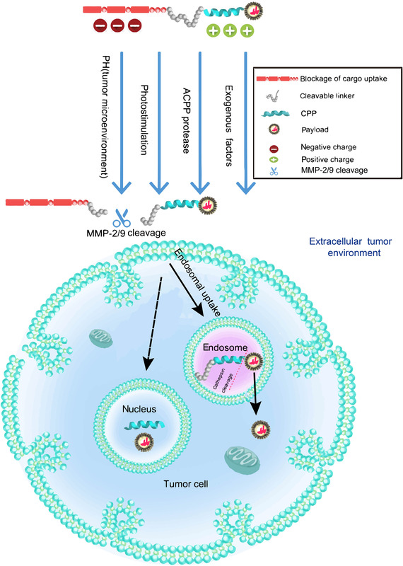
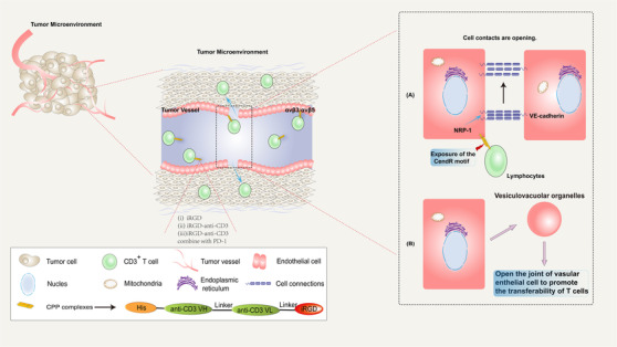
Due to the complex physiological structure, microenvironment and multiple physiological barriers, traditional anti-cancer drugs are severely restricted from reaching the tumour site. Cell-penetrating peptides (CPPs) are typically made up of 5-30 amino acids, and can be utilised as molecular transporters to facilitate the passage of therapeutic drugs across physiological barriers. Up to now, CPPs have widely been used in many anti-cancer treatment strategies, serving as an excellent potential choice for oncology treatment. However, their drawbacks, such as the lack of cell specificity, short duration of action, poor stability in vivo, compatibility problems (i.e. immunogenicity), poor therapeutic efficacy and formation of unwanted metabolites, have limited their further application in cancer treatment. The cellular uptake mechanisms of CPPs involve mainly endocytosis and direct penetration, but still remain highly controversial in academia. The CPPs-based drug delivery strategy could be improved by clever design or chemical modifications to develop the next-generation CPPs with enhanced cell penetration capability, stability and selectivity. In addition, some recent advances in targeted cell penetration that involve CPPs provide some new ideas to optimise CPPs.

Keywords: Anti-cancer therapy; cell-penetrating peptides; molecular cargoes; optimisation; tumour immunity.


Figures

FIGURE 1
FIGURE 1
Schematic diagram illustrating the types of CPPs. CPPs are classified in different ways: (A) CPPs can be classified as D type, L type, mix type and modified type based on chirality or modification. (B) According to the conformation, linear peptides are in the majority. (C) CPPs may be derived from different sources, with synthetic CPPs accounting for the largest part. (D) CPPs can be utilised as DDSs and be divided into different subgroups based on their cargoes. Specific classification results and their proportions have been marked in the above figure. The most popular classification is according to physical–chemical properties, in which CPPs can be classified into three subgroups: cationic CPPs, amphipathic CPPs and hydrophobic CPPs
FIGURE 2
FIGURE 2
Chronological arrow in CPP development. Those in the blue box represent important CPPs with epoch‐making significance, and those in yellow are major events. ATTEMPT, ACPP and tumour homing CPPs can be seen in Part 4
FIGURE 3
FIGURE 3
Mechanisms of the intracellular entry of CPPs. Schematic representation of mechanisms for CPPs internalisation. The involved pathways can be divided into two groups: endocytosis (blue) and energy‐independent mechanisms (pink). Endocytosis pathways consist of macropinocytosis, caveolin‐mediated endocytosis, clathrin‐mediated endocytosis and clathrin and caveolin‐independent endocytosis. Energy‐independent mechanisms have been proposed to occur through: the ‘barrel‐stave’ model, the ‘carpet‐like’ model, the inverted micelle model, the membrane thinning mode and another hypothesis of ‘membrane thinning’. The small molecules involved in the uptake progress have been marked in the domain of the relevant pathway
FIGURE 4
FIGURE 4
Mechanisms to enhance the specialty of CPPs. Several schematic diagrams of CPP compounds to improve the specificity: (A) The polycation active domain is cationic and the shielding domain is anionic. The cleavable linking arm is the key to specificity. (B) The cationic peptide allows selective entry of the cargoes into cells. (C) The combination of CPPs with the antibody has been proved to improve the specificity. (D) Works by targeting receptors or ligands. (E) EPR refers to the enhanced permeability and retention effect. Passive targeting based on EPR can be utilised to improve the specificity of CPPs
FIGURE 5
FIGURE 5
The activation and cellular uptake of ACPPs. ACPPs are a new type of carrier that can be activated by special enzymes in the tumour tissue site to induce cell penetration. The molecular structure generally includes three functional regions: polycation active domain with the cell‐penetrating ability (e.g. CPPs); a cleavable connecting arm; polyanion shielding domain. As shown in the figure, there is the high‐expressed protease at the tumour site. Certain internal environmental factors such as low pH, and external physicochemical stimulation such as light and exogenous substances are the conditions that can dependently trigger cleavage between polycation active domain and polyanion shielding domain. This schematic diagram selects the most potential protease MMP2/9 as a shear to activate the activatable CPP compound. After cleavage in specific sites, they can enter tumour cells by uptake mechanisms. The dashed arrow means the entrance into the nucleus is sometimes observed, and the mechanism is not fully understood
FIGURE 6
FIGURE 6
Mechanisms of CPP act in immunotherapy. Circulating iRGD‐modified T cells are tethered and rolled in blood flow by the engagement of αvβ3/αvβ5 expressed on tumour vascular endothelial cells. There are three CPP complexes: (i) IRGD (Lowest curative effect); (ii) iRGD‐anti‐CD3 (Medium curative effect) and (iii) iRGD‐anti‐CD3 combine with PD‐1. (Best curative effect) The key to facilitating tumour infiltration is to promote the opening of cell connections. There are two main mechanisms: (A) The interaction also initiates the proteolysis of iRGD and expose the CendR motif. The truncated peptide then binds to NRP‐1, triggering the tyrosine phosphorylation of VE‐cadherin and the formation of intercellular gaps. (B) Another vesicular transport pathway in the endothelial cytoplasm is termed vesiculovacuolar organelles. Then, the connected lymphocytes cross the vessel wall and infiltrate into the tumour parenchyma

Similar articles

Cited by

References

    1. Kardani K, Milani A, Shabani HS, Bolhassani A. Cell penetrating peptides: the potent multi‐cargo intracellular carriers. Expert Opin Drug Deliv. 2019;16(11):1227‐1258. 10.1080/17425247.2019.1676720. - DOI - PubMed
    1. Heitz F, Morris MC, Divita G. Twenty years of cell‐penetrating peptides: from molecular mechanisms to therapeutics. Br J Pharmacol. 2009;157(2):195‐206. 10.1111/j.1476-5381.2009.00057.x. - DOI - PMC - PubMed
    1. Guidotti G, Brambilla L, Rossi D. Cell‐penetrating peptides: from basic research to clinics. Trends Pharmacol Sci. 2017;38(4):406‐424. 10.1016/j.tips.2017.01.003. - DOI - PubMed
    1. Frankel AD, Pabo CO. Cellular uptake of the tat protein from human immunodeficiency virus. Cell. 1988;55(6):1189‐1193. 10.1016/0092-8674(88)90263-2. - DOI - PubMed
    1. Schwarze SR, Ho A, Vocero‐Akbani A, Dowdy SF. In vivo protein transduction: delivery of a biologically active protein into the mouse. Science (New York, N.Y.). 1999;285(5433):1569‐1572. 10.1126/science.285.5433.1569. - DOI - PubMed

Publication types

Substances