. 2022 Jun 1;15(6):dmm049544.
doi: 10.1242/dmm.049544. Epub 2022 Jun 23.

Snrpb is required in murine neural crest cells for proper splicing and craniofacial morphogenesis

Affiliations

Snrpb is required in murine neural crest cells for proper splicing and craniofacial morphogenesis

Sabrina Shameen Alam et al. Dis Model Mech. .

Abstract

Heterozygous mutations in SNRPB, an essential core component of the five small ribonucleoprotein particles of the spliceosome, are responsible for cerebrocostomandibular syndrome (CCMS). We show that Snrpb heterozygous mouse embryos arrest shortly after implantation. Additionally, heterozygous deletion of Snrpb in the developing brain and neural crest cells models craniofacial malformations found in CCMS, and results in death shortly after birth. RNAseq analysis of mutant heads prior to morphological defects revealed increased exon skipping and intron retention in association with increased 5' splice site strength. We found increased exon skipping in negative regulators of the P53 pathway, along with increased levels of nuclear P53 and P53 target genes. However, removing Trp53 in Snrpb heterozygous mutant neural crest cells did not completely rescue craniofacial development. We also found a small but significant increase in exon skipping of several transcripts required for head and midface development, including Smad2 and Rere. Furthermore, mutant embryos exhibited ectopic or missing expression of Fgf8 and Shh, which are required to coordinate face and brain development. Thus, we propose that mis-splicing of transcripts that regulate P53 activity and craniofacial-specific genes contributes to craniofacial malformations. This article has an associated First Person interview with the first author of the paper.

Keywords: SNRPB; Cerebrocostomandibular syndrome; Craniofacial; Neural crest cells; Splicing.


Conflict of interest statement

Competing interests The authors declare no competing or financial interests.

Figures
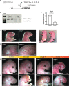
Fig. 1.
Fig. 1.
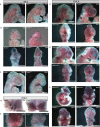
Deletion of exons 2-3 of Snrpb results in reduced Snrpb level and craniofacial malformations of varying severity. (A) Schema of the conditional SnrpbloxP/+ allele generated using CRISPR/Cas9 and the deletion generated in the presence of Cre. (B) Deletion of the loxP-flanked exons 2-3 in Snrpb produces a shorter transcript of 527 bp. (C) Quantitative RT-PCR showing a significant decrease in Snrpb level in E8.5 heterozygous mutant embryos (**P<0.01, unpaired, two-tailed t-test). (D) P0 Snrpbncc+/− (SnrpbloxP/+; Wnt-1Cre2tg/+) pups have an abnormally shaped head, micrognathia and abnormal outer ears. (E) Higher-magnification images showing a hypoplastic pinna (E) in the Snrpbncc+/− mutant (black arrow) compared to control (SnrpbL/+). (F) Snrpbncc+/− mutant pups lack a milk spot in their stomach (black arrowhead), which is visible in the Snrpb wild-type (WT) littermate (white arrowhead). (G,H) Representative images of Snrpb WT (SnrpbL/+) and Snrpbncc+/− mutant embryos at E14.5 (G) and E17.5 (H). Snrpbncc+/− mutant embryos show a range of craniofacial malformations: Group 2 had abnormal outer ear, and cranial and mandibular hypoplasia; Group 3 had nasal clefts; Group 4 showed severe abnormalities including absence of the head and face. AU, arbitrary units; E, ear; Fl, forelimb; M, mandible; N, nose; Y, eye. Scale bars: 1 mm.
Fig. 2.
Fig. 2.
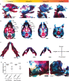
Craniofacial cartilage and bones of Snrpbncc+/− embryos are hypoplastic or missing. (A,B) Representative images of Alcian Blue- and Alizarin Red-stained E17.5 normal (Snrpbwt or SnrpbL/+) and Snrpbncc+/− mutant embryos showing craniofacial abnormalities of varying penetrance. (A) Sagittal view showing hypoplasia or absence of neural crest cell-derived bones (labeled in green font) in Snrpbncc+/− mutants. The missing hyoid bone and tracheal cartilage are indicated by red arrows. (B) Ventral view of the skull showing palatal and maxillary clefts (stars) in group 2 and 3 Snrpbncc+/− mutants, respectively, and the absence of the ventral craniofacial components in group 4 mutants. (C) Representative images of the lower jaw of a normal embryo and two Snrpbncc+/− mutants, showing asymmetric mandibles with no discernable angular, coronoid or condylar processes. (D) Both left and right mandibles are significantly shorter in Snrpbncc+/− embryos (***P<0.0001, unpaired, two-tailed t-test), compared to controls (Snrpbwt and SnrpbL/+ embryos). (E) Representative higher-magnification images of the inner ear of a control and Snrpbncc+/− embryo. Middle ear structures such as the stapes were absent in group 3 and 4 mutants, whereas a presumably duplicated Meckel's cartilage and ectopic structures were found in a subset. an, angular process; As, alisphenoid bone; bo, basioccipital bone; bs, basisphenoid bone; cn, condylar process; cp, coronoid process; fb, frontal bone; hb, hyoid bone; in, incus; ip, intraparietal bone; mc, Meckel's cartilage; ml, malleus; mn, mandible; MX, maxilla; nb, nasal bone; nc, nasal cartilage; ob, occipital bone; pb, parietal bone; PMX, premaxilla; pl, palatine; PPMX, palatal process of maxilla; Sq, squamous bone; st, stapes; Za, zygomatic arch. Scale bars: 500 μm.
Fig. 3.
Fig. 3.
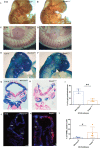
Abnormal cranial ganglia, reduced cranial neural crest cells and a significant increase in cell death in Snrpbncc+/− mutants. (A,B) Representative images of E10.5 control (SnrpbL/+) (A) and Snrpbncc+/− (B) group 2 embryos stained with antibody against neurofilament (2H3). (B) Snrpbncc+/− mutants (n=2) showed abnormal projections of nerves to the pharyngeal arches (cranial ganglion v and vii) and heart (cranial ganglion ix), and absence and abnormal bundling of cranial nerves (all are indicated by black arrowheads). (C,D) Compared to controls (SnrpbL/+) (C), the dorsal root ganglia are bifurcated and reduced in mutants (black arrowheads) (D). (E-H) Representative images of X-gal-stained wholemount (E,F) and sectioned (G,H) E10.5 control (Wnt-Cre2tg/+) (E,G) and Snrpbncc+/− group 2 mutant (F,H) embryos. Snrpbncc+/− mutants show reduced X-gal staining in the craniofacial region and in the pharyngeal arches (n=4) compared to the control embryos (n=4). (I) Quantification of the area stained with X-gal showed a significant reduction in mutants (n=3) compared to control littermates (n=3) (unpaired, two-tailed t-test, **P<0.005). Error bars indicate s.e.m. (J,K) Representative images of sections of TUNEL-stained E9.5 control (Snrpbwt or SnrpbL/+) (J) and Snrpbncc+/− mutant (K) embryos. (L) Quantification showed an increase in the percentage of TUNEL-positive nuclei (red in J,K) in the craniofacial region of mutants (n=3) (unpaired, two-tailed t-test, *P<0.05). Error bars indicate s.e.m. 1, 2, 3, pharyngeal arches 1, 2 and 3; drg, dorsal root ganglia; fb, forebrain; fl, forelimb; hb, hindbrain; hm, head mesenchyme; Ht, heart; mb, midbrain; mx, maxillary prominence; nt, neural tube; pa, pharyngeal arch. Scale bars: 500 μm (A-H), 50 μm (J,K).
Fig. 4.
Fig. 4.
Snrpbncc+/− mutant heads show aberrant splicing including increased exon skipping and intron retention. (A) Differentially expressed genes (DEGs) identified in Snrpbncc+/− (Het) mutants could be grouped into molecular pathways belonging to the P53 signaling pathway and the spliceosome. (B) A much larger number of transcripts was found to be abnormally spliced in Snrpbncc+/− mutants compared to controls (Snrpbwt). The most abundant differentially spliced events (DSEs) were skipped exons (SEs) and retained introns (RIs). A strong tendency towards increased exon skipping and intron inclusion in the mutant samples was observed; there were more SEs (273 in Het versus 83 in WT) and RIs (191 in Het versus 21 in WT). DSEs were predicted to introduce premature termination codons (PTCs) more often in Hets compared to WT. (C-F) In Hets, exon skipping was significantly higher for non-constitutive exons (NonCE) when constitutive (CE) versus (NonCE) was examined (unpaired, two-tailed t-test). (G-J) An analysis of splice site (SS) strengths in DSEs revealed significantly stronger 5′ SS in mutants than in controls (P=0.05, unpaired, two-tailed t-test). (K) Pathway analysis of genes with DSEs showed that they were strongly associated with mRNA processing. ns, not significant; *P<0.05, **P<0.01.
Fig. 5.
Fig. 5.
Increased exon skipping in two regulators of P53, Mdm2 and Mdm4, and significant increases in P53 target genes in Snrpbncc+/− mutant heads. (A,B) Representative images of RT-PCR showing long (FL) and short transcripts (ΔE) produced in Mdm2 (A) and Mdm4 (B) in E9.0 control (Snrpbwt or SnrpbL/+) and Snrpbncc+/− embryos. Quantification revealed a significant increase in the ratio of Mdm2 and Mdm4 transcripts containing a skipped exon over the longer transcript in mutants (unpaired, two-tailed t-test, *P<0.05). Error bars indicate s.d. (C) Levels of P53 target genes were significantly increased in morphologically normal E9.0 Snrpbncc+/− embryos (n=5) compared to controls (n=5) (unpaired, two-tailed t-test, *P<0.05). (D) At E9.5, although levels of P53 target genes were increased in Snrpbncc+/− embryos (n=3 group 1 and n=2 group 2), this difference was not significant. Error bars indicate s.e.m. FL, full length transcript; ΔE3, transcript with exon 3 skipped; ΔE7, transcript with exon 7 skipped.
Fig. 6.
Fig. 6.
Transcripts with exon skipping events are found in heads of E9.0 control and mutant embryos. (A-C) Sashimi plots for the exon skipping events found for Smad2 (A), Pou2f1 (B) and Rere (C). Under each sashimi plot, representative gel for RT-PCR showing the presence of transcripts with the predicted exon skipping event. The location of primers used to amplify transcripts is shown on the right. No significant difference was found in the ratio of short/long transcripts (unpaired, two-tailed t-test). Error bars in the graphs indicate s.e.m. FL, full length; ΔE, skipped exon.
Fig. 7.
Fig. 7.
Shh, Fgf8 and Msx2 are mis-expressed in Snrpbncc+/− embryos. Representative images of E9.5 and E10.5 control (Wnt-Cre2Tg/+ or Snrpbwt) and Snrpbncc+/− embryos after wholemount in situ hybridization (ISH) to detect mRNA expression of Shh, Fgf8 and Msx2. (A) Shh at E9.5 was expressed in the ventral-most region of the neural tube, the floor plate, and the ventral prosencephalon of control and Snrpbncc+/− embryos (black arrowheads). (B) Lateral (top row) and ventral (bottom row) views of embryos showing that expression of Fgf8 was expanded in the mandibular epithelium, the frontonasal prominence and the midbrain/hindbrain junction in group 2 (black arrowheads) (n=4) mutant embryos. (C) Msx2 expression was extended proximally in pharyngeal arches 1 and 2 in group 1 mutant embryos (n=2; black arrowheads). (D) Wholemount ISH at E10.5. Top row: lateral view of embryos showing Shh expression in the diencephalon and ventral forebrain in a control (left) and group 2 mutant (right) embryo. Ectopic expression was found in the dorsal and ventral optic lens (arrowheads) of the mutant. Bottom row: ventral view showing expression of Shh on the oral ectoderm in a control (left) and group 2 mutant (right) embryo (black arrowheads). (E) Top row shows representative images of lateral views of a control embryo, in which Fgf8 was detected on the surface ectoderm of the mandible, maxillary and frontonasal prominences (n=3). In the representative group 2 Snrpbncc+/− mutant embryo, Fgf8 expression was observed on the mandibular ectoderm and in the region where the maxillary prominence would normally form (black arrowheads; n=3); ectopic expression was also detected in the lens. Bottom row shows frontal view of reduced Fgf8 expression in the hypoplastic lateral nasal prominence in mutants, and ectopic expression of Fgf8 on the surface ectoderm of the medial nasal process, towards the midline (black arrowheads). (F) Top row shows a lateral (left) and ventral (right) view of Msx2 expression in E10.5 control embryos. Bottom row shows that in E10.5 group 1 Snrpbncc+/− mutants on the left, Msx2 was expressed in the lateral and medial nasal prominences, although expression appeared reduced and ventrally expanded in the medial frontal nasal region. On the right, in a group 2 Snrpbncc+/− mutant with a hypoplastic frontonasal and maxillary prominences, Msx2 was expressed in the region where the maxillary prominences would form and in the mandibular region of the hypoplastic first arch. (G,H) Higher-magnification images of the facial ectoderm zone (FEZ) region of E10.5 embryos showing Shh expression in the developing mandibular periderm, and bilaterally on the surface ectoderm of the medial nasal prominences of control (G,H, left) and group 2 Snrpbncc+/− (G,H, right) embryos (black arrowheads). (H) Fgf8 expression in the lateral nasal prominence was reduced in group 2 mutants (yellow arrowhead), while ectopic expression of Fgf8 was found on the surface ectoderm of the medial nasal process, towards the midline. 1, 2, pharyngeal arches 1 and 2; FNP, frontonasal process; HB, hindbrain; HT, heart; MB, midbrain; MP, mandibular process; NP, nasal process; OR, optic region. Scale bars: 500 μm.

Similar articles

Cited by

References

    1. Anders, S., Pyl, P. T. and Huber, W. (2015). HTSeq--a Python framework to work with high-throughput sequencing data. Bioinformatics 31, 166-169. 10.1093/bioinformatics/btu638 - DOI - PMC - PubMed
    1. Bacrot, S., Doyard, M., Huber, C., Alibeu, O., Feldhahn, N., Lehalle, D., Lacombe, D., Marlin, S., Nitschke, P., Petit, F.et al. (2015). Mutations in SNRPB, encoding components of the core splicing machinery, cause cerebro-costo-mandibular syndrome. Hum. Mutat. 36, 187-190. 10.1002/humu.22729 - DOI - PubMed
    1. Beauchamp, M. C., Alam, S. S., Kumar, S. and Jerome-Majewska, L. A. (2020). Spliceosomopathies and neurocristopathies: Two sides of the same coin? Dev. Dyn. 249, 924-945. 10.1002/dvdy.183 - DOI - PubMed
    1. Beauchamp, M.-C., Djedid, A., Bareke, E., Merkuri, F., Aber, R., Tam, A. S., Lines, M. A., Boycott, K. M., Stirling, P. C., Fish, J. L.et al. (2021). Mutation in Eftud2 causes craniofacial defects in mice via mis-splicing of Mdm2 and increased P53. Hum. Mol. Genet. 30, 739-757. 10.1093/hmg/ddab051 - DOI - PMC - PubMed
    1. Bogue, M. A., Grubb, S. C., Walton, D. O., Philip, V. M., Kolishovski, G., Stearns, T., Dunn, M. H., Skelly, D. A., Kadakkuzha, B., TeHennepe, G.et al. (2018). Mouse Phenome Database: an integrative database and analysis suite for curated empirical phenotype data from laboratory mice. Nucleic Acids Res. 46, D843-D850. 10.1093/nar/gkx1082 - DOI - PMC - PubMed

Publication types

Supplementary concepts

Grants and funding