

Identification of PBK as a hub gene and potential therapeutic target for medulloblastoma
- PMID: 35593307
- PMCID: PMC9164263
- DOI: 10.3892/or.2022.8336
Identification of PBK as a hub gene and potential therapeutic target for medulloblastoma
Abstract
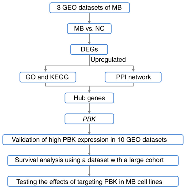
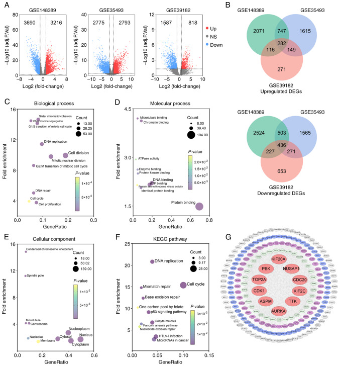
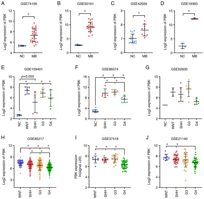
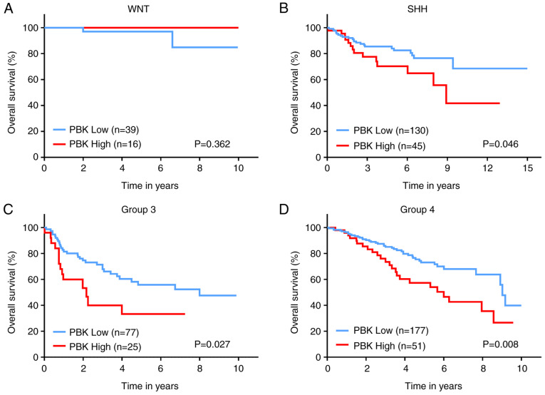
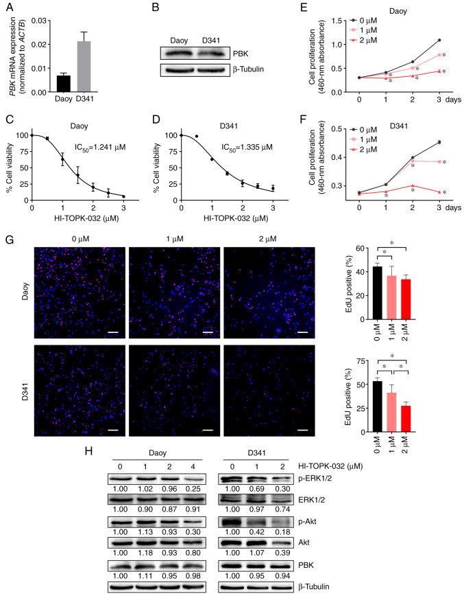
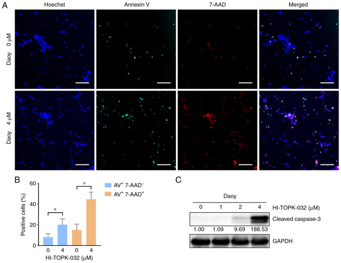
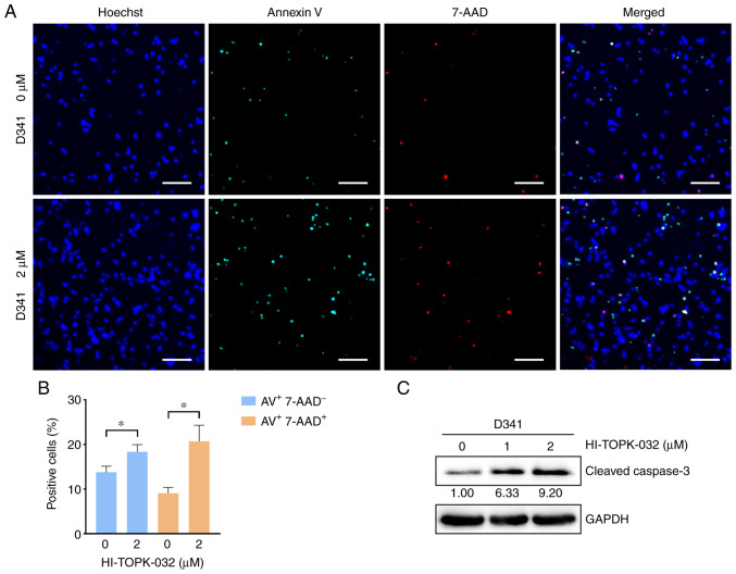
Medulloblastoma (MB) is the most frequent malignant brain tumor in pediatrics. Since the current standard of care for MB consisting of surgery, cranio‑spinal irradiation and chemotherapy often leads to a high morbidity rate, a number of patients suffer from long‑term sequelae following treatment. Targeted therapies hold the promise of being more effective and less toxic. Therefore, the present study aimed to identify hub genes with an upregulated expression in MB and to search for potential therapeutic targets from these genes. For this purpose, gene expression profile datasets were obtained from the Gene Expression Omnibus database and processed using R 3.6.0 software to screen differentially expressed genes (DEGs) between MB samples and normal brain tissues. A total of 282 upregulated and 436 downregulated DEGs were identified. Functional enrichment analysis revealed that the upregulated DEGs were predominantly enriched in the cell cycle, DNA replication and cell division. The top 10 hub genes were identified from the protein‑protein interaction network of upregulated genes, and one identified hub gene [PDZ binding kinase (PBK)] was selected for further investigation due to its possible role in the pathogenesis of MB. The aberrant expression of PBK in MB was verified in additional independent gene expression datasets. Survival analysis demonstrated that a higher expression level of PBK was significantly associated with poorer clinical outcomes in non‑Wingless MBs. Furthermore, targeting PBK with its inhibitor, HI‑TOPK‑032, impaired the proliferation and induced the apoptosis of two MB cell lines, with the diminished phosphorylation of downstream effectors of PBK, including ERK1/2 and Akt, and the activation of caspase‑3. Hence, these results suggest that PBK may be a potential prognostic biomarker and a novel candidate of targeted therapy for MB.
Keywords: PDZ binding kinase; apoptosis; medulloblastoma; proliferation; therapeutic target.
Conflict of interest statement
The authors declare that they have no competing interests.
Figures
Similar articles
-
The LIN28B-let-7-PBK pathway is essential for group 3 medulloblastoma tumor growth and survival.Mol Oncol. 2023 Sep;17(9):1784-1802. doi: 10.1002/1878-0261.13477. Epub 2023 Aug 7. Mol Oncol. 2023. PMID: 37341142 Free PMC article.
-
Identification of Potential Key Genes and Molecular Mechanisms of Medulloblastoma Based on Integrated Bioinformatics Approach.Biomed Res Int. 2022 Jan 4;2022:1776082. doi: 10.1155/2022/1776082. eCollection 2022. Biomed Res Int. 2022. Retraction in: Biomed Res Int. 2024 Mar 20;2024:9853643. doi: 10.1155/2024/9853643. PMID: 35127939 Free PMC article. Retracted.
-
Bioinformatics analyses of significant genes, related pathways and candidate prognostic biomarkers in glioblastoma.Mol Med Rep. 2018 Nov;18(5):4185-4196. doi: 10.3892/mmr.2018.9411. Epub 2018 Aug 21. Mol Med Rep. 2018. PMID: 30132538 Free PMC article.
-
Identification of ITPR1 as a Hub Gene of Group 3 Medulloblastoma and Coregulated Genes with Potential Prognostic Values.J Mol Neurosci. 2022 Mar;72(3):633-641. doi: 10.1007/s12031-021-01942-3. Epub 2021 Nov 25. J Mol Neurosci. 2022. PMID: 34822110
-
Molecular Classification of Medulloblastoma.Neurol Med Chir (Tokyo). 2016 Nov 15;56(11):687-697. doi: 10.2176/nmc.ra.2016-0016. Epub 2016 May 26. Neurol Med Chir (Tokyo). 2016. PMID: 27238212 Free PMC article. Review.
Cited by
-
The pivotal role of irradiation-induced apoptosis in the pathogenesis and therapy of medulloblastoma.Cancer Rep (Hoboken). 2024 Apr;7(4):e2048. doi: 10.1002/cnr2.2048. Cancer Rep (Hoboken). 2024. PMID: 38599791 Free PMC article. Review.
-
The LIN28B-let-7-PBK pathway is essential for group 3 medulloblastoma tumor growth and survival.Mol Oncol. 2023 Sep;17(9):1784-1802. doi: 10.1002/1878-0261.13477. Epub 2023 Aug 7. Mol Oncol. 2023. PMID: 37341142 Free PMC article.
-
Comparative Efficacy of Tumor Microenvironment-responsive Nanotherapeutics Targeting PSD95/Discs-large/ZO-1 Binding Kinase in Different Histological Subgroups of Medulloblastoma.Int J Med Sci. 2024 Nov 11;21(15):3018-3033. doi: 10.7150/ijms.97992. eCollection 2024. Int J Med Sci. 2024. PMID: 39628686 Free PMC article.
References
MeSH terms
Substances
LinkOut - more resources
Full Text Sources
Research Materials
Miscellaneous
