

Bone Marrow Lesions and Magnetic Resonance Imaging-Detected Structural Abnormalities in Patients With Midfoot Pain and Osteoarthritis: A Cross-Sectional Study
- PMID: 35593411
- PMCID: PMC10952448
- DOI: 10.1002/acr.24955
Bone Marrow Lesions and Magnetic Resonance Imaging-Detected Structural Abnormalities in Patients With Midfoot Pain and Osteoarthritis: A Cross-Sectional Study
Abstract
Objective: To compare magnetic resonance imaging (MRI)-detected structural abnormalities in patients with symptomatic midfoot osteoarthritis (OA), patients with persistent midfoot pain, and asymptomatic controls, and to explore the association between MRI features, pain, and foot-related disability.
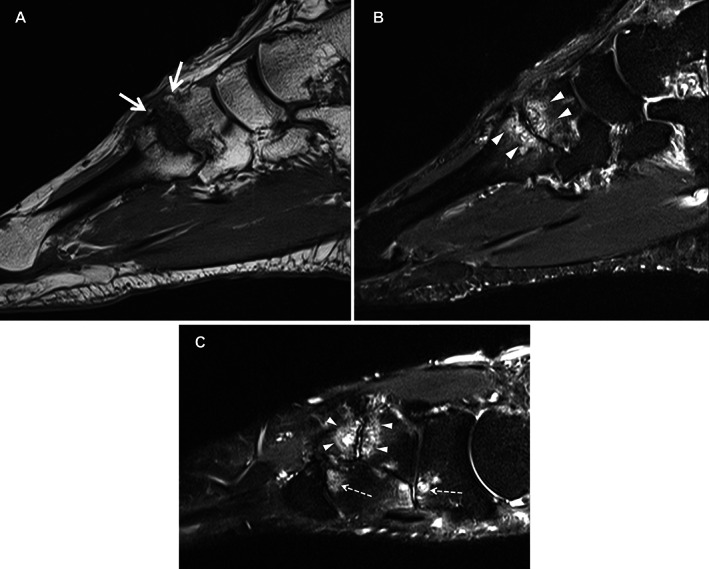
Methods: One hundred seven adults consisting of 50 patients with symptomatic and radiographically confirmed midfoot OA, 22 adults with persistent midfoot pain but absence of radiographic OA, and 35 asymptomatic adults underwent 3T MRI of the midfoot and clinical assessment. MRIs were read for the presence and severity of abnormalities (bone marrow lesions [BMLs], subchondral cysts, osteophytes, joint space narrowing [JSN], effusion-synovitis, tenosynovitis, and enthesopathy) using the Foot Osteoarthritis MRI Score. Pain and foot-related disability were assessed with the Manchester Foot Pain and Disability Index.
Results: The severity sum score of BMLs in the midfoot was greater in patients with midfoot pain and no signs of OA on radiography compared to controls (P = 0.007), with a pattern of involvement in the cuneiform-metatarsal joints similar to that in patients with midfoot OA. In univariable models, BMLs (ρ = 0.307), JSN (ρ = 0.423), and subchondral cysts (ρ = 0.302) were positively associated with pain (P < 0.01). In multivariable models, MRI abnormalities were not associated with pain and disability when adjusted for covariates.
Conclusion: In individuals with persistent midfoot pain but no signs of OA on radiography, MRI findings suggested an underrecognized prevalence of OA, particularly in the second and third cuneiform-metatarsal joints, where BML patterns were consistent with previously recognized sites of elevated mechanical loading. Joint abnormalities were not strongly associated with pain or foot-related disability.
© 2022 The Authors. Arthritis Care & Research published by Wiley Periodicals LLC on behalf of American College of Rheumatology.
Figures


References
-
- Golightly YM, Gates LS. Foot osteoarthritis: addressing an overlooked global public health problem. Arthritis Care Res (Hoboken) 2021;73:767–9. - PubMed
-
- Rao S, Baumhauer J, Nawoczenski D. Is barefoot regional plantar loading related to self‐reported foot pain in patients with midfoot osteoarthritis. Osteoarthritis Cartilage 2011;19:1019–25. - PubMed
Publication types
MeSH terms
Grants and funding
LinkOut - more resources
Full Text Sources
Medical
