. 2022 Jun 1;135(11):jcs259617.
doi: 10.1242/jcs.259617. Epub 2022 May 31.

VEGF-A controls the expression of its regulator of angiogenic functions, dopamine D2 receptor, on endothelial cells

Affiliations

VEGF-A controls the expression of its regulator of angiogenic functions, dopamine D2 receptor, on endothelial cells

Chandrani Sarkar et al. J Cell Sci. .

Abstract

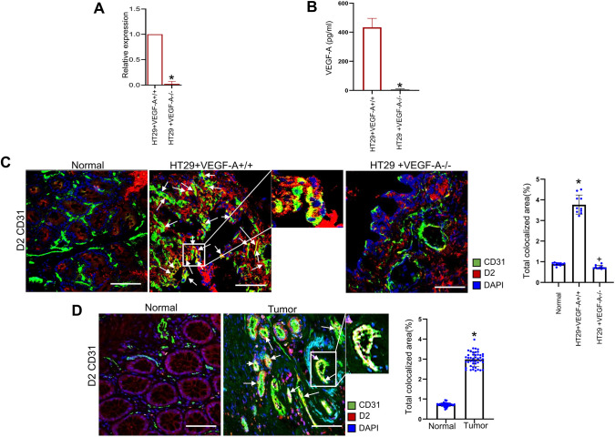
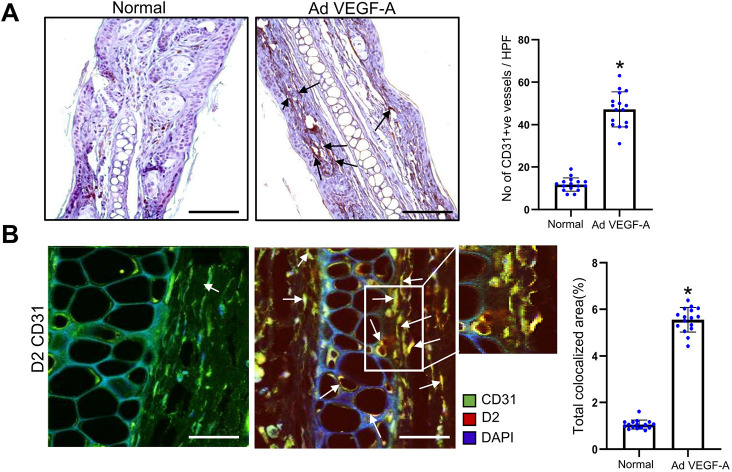
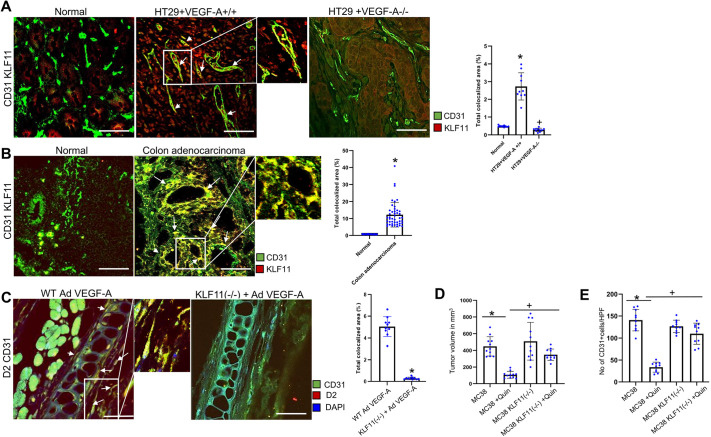
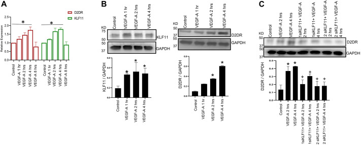
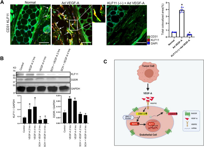
We have previously demonstrated significant upregulation of dopamine D2 (DAD2) receptor (DRD2) expression on tumor endothelial cells. The dopamine D2 receptors, upon activation, inhibit the proangiogenic actions of vascular endothelial growth factor-A (VEGF-A, also known as vascular permeability factor). Interestingly, unlike tumor endothelial cells, normal endothelial cells exhibit very low to no expression of dopamine D2 receptors. Here, for the first time, we demonstrate that through paracrine signaling, VEGF-A can control the expression of dopamine D2 receptors on endothelial cells via Krüppel-like factor 11 (KLF11)-extracellular signal-regulated kinase (ERK) 1/2 pathway. These results thus reveal a novel bidirectional communication between VEGF-A and DAD2 receptors.

Keywords: Dopamine D2 receptor; Endothelial cell; VEGF-A.


Conflict of interest statement

Competing interests The authors declare no competing or financial interests.

Figures

Fig. 1.
Fig. 1.
VEGF-A affects DAD2 receptor expression in endothelial cells. Knockdown of VEGF-A in HT29 cells was confirmed by (A) real-time qPCR and (B) ELISA. Data are expressed as the mean±s.e.m. (n=3). *P<0.05 (two-sample two-tailed t-test). (C) Representative images of colocalization (arrows) of CD31 and DAD2 receptors [i.e. expression of DAD2 receptors on the tumor endothelial cells (TECs)]. TECs (green) of HT29+VEGF-A+/+ tumors showed significantly higher expressions of DAD2 receptors (red) compared to TECs of HT29+VEGF-A/ tumors and endothelial cells (ECs) of normal colon tissues (n=10 for each group). Scale bars: 80 µm. Right, ten fields of view were randomly chosen and analyzed. Data are expressed as the mean±s.e.m. *P<0.05 for HT29+VEGF-A+/+ versus normal; +P<0.05 for HT29+VEGF-A/ versus HT29+VEGF-A+/+ (one-way ANOVA with Bonferroni step down adjustment). (D) Representative images of colocalization (arrows) of CD31/dopamine D2 receptors indicate high expressions of DAD2 receptors on TECs of VEGF-A secreting well-differentiated human colon adenocarcinoma tissues compared to the ECs of normal colon tissues (n=45 for each group). Scale bars: 80 µm. Right, four fields of view per section were randomly chosen and analyzed. Data are expressed as the mean±s.e.m. *P<0.05 for tumor versus normal (two-sample two-tailed t-test).
Fig. 2.
Fig. 2.
High DAD2 receptor expression is observed in the endothelial cells upon AdVEGF-A injection. (A) Representative images of immunohistochemistry (arrows) showing increased angiogenesis (CD31 expression) in the AdVEGF-A-treated ear of the C57/Bl6 mouse compared to the vehicle-treated control ear of the same mouse (n=16 for each group. Scale bars: 100 µm. Right, ten randomly chosen fields were used to quantify the microvessel numbers, which is expressed as the number of microvessels/high power fields. Data are expressed as mean±s.e.m. *P<0.005 AdVEGF-A relative to vehicle-treated control (two-sample two-tailed t-test). (B) Representative images of colocalization (arrows) of CD31 and DAD2 receptors indicating higher expression of DAD2 receptors (red) on the endothelial cells (green) in AdVEGF-A-injected ears compared to endothelial cells of vehicle-treated normal ear blood vessels (n=16 for each group). Scale bars: 40 µm. Right, ten fields of view were randomly chosen and analyzed. Data are expressed as mean±s.e.m. *P<0.05 for AdVEGF-A relative to vehicle-treated control (two-sample two-tailed t-test).
Fig. 3.
Fig. 3.
VEGF-A induces KLF11 in endothelial cells. (A) Representative images of colocalization (arrows) of CD31 and KLF11 indicating significantly higher expression of KLF11 (red) in CD31-positive vessels (green) in VEGF-A-secreting HT29 tumors than CD31-positive vessels of normal colon tissues and HT29 +VEGF-A−/− tumors (n=10 for each group). Scale bars: 80 µm. Right, ten fields of view were randomly chosen and analyzed. Data are expressed as the mean±s.e.m. *P<0.05 for HT29+VEGF+/+ compared with the normal; +P<0.05 HT29+VEGF-A−/− compared with HT29+VEGF-A+/+ (one-way ANOVA with Bonferroni step down adjustment). (B) Representative images of colocalization (arrows) of CD31 and KLF11, indicating higher KLF11 expression (red) in tumor endothelial cells (TEC) (green) of VEGF-A-secreting well-differentiated colon adenocarcinoma tissues than on endothelial cells (green) of normal colon tissues (n=45 each group). Scale bars: 80 µm. Right, four fields of view per section were randomly chosen and analyzed. Data are expressed as the mean±s.e.m. *P<0.05 for colon adenocarcinoma compared with the normal (two-sample two-tailed t-test). (C) Colocalization (arrows) of CD31 and DAD2 receptors showing that AdVEGF-A injection into ears of wild-type (WT) C57/Bl6 mice resulted in upregulation of DAD2 receptors (red) in the endothelial cells (green) of ear blood vessels of these mice. However, AdVEGF-A injection into the ears of endothelial cell-specific KLF11(−/−) mice failed to lead to any significant upregulation in the expression of DAD2 receptors in the endothelial cells of ear blood vessels (n=10 in each group). Scale bars: 40 µm. Right, ten fields of view were randomly chosen and analyzed. Data are expressed as the mean±s.e.m. *P<0.05 for KLF11 (−/−) compared with wild-type C57/Bl6 mice injected with AdVEGF-A (two-sample two-tailed t-test). (D) DAD2 receptor agonist quinpirole (10 mg/kg of body weight intraperitoneally once daily for 7 days) significantly reduces MC38 tumor growth and (E) the number of CD31-positive cells [i.e. microvessel density (MVD) or angiogenesis]. However, inhibition by quinpirole was significantly more in tumor-bearing wild-type C57BL6 mice than in endothelial cell-specific KLF11(−/−) C57BL6 mice bearing MC38 colon tumors. Data are expressed as mean±s.e.m. *P<0.005 when compared to control MC38; +P<0.005 when compared to MC38+Quin (n=11 each group) (one-way ANOVA with Bonferroni step down adjustment). HPF, high power field of view.
Fig. 4.
Fig. 4.
KLF11 and D2DR expression in HUVECs upon treatment with VEGF-A. (A) Real-time qPCR indicates a time-dependent increase in KLF11 expression in HUVECs upon VEGF-A treatment (100 ng/ml of human recombinant VEGF-A) with maximum expression at 4 h. A similar increase was also observed in D2DR expression in HUVECs upon VEGF-A treatment. Data are expressed as mean±s.e.m., n=3 (relative to vehicle-treated control). *P<0.0001 (one-way ANOVA with Bonferroni step down adjustment). (B) Western blot analysis showing significant upregulation of KLF11 expression within 1 h of stimulation and DAD2 receptor (D2DR) expression within 2 h of stimulation in HUVECs treated with 100 ng/ml of human recombinant VEGF-A. The upregulation was also evident at 4 h. The fold changes of KLF11 and D2DR normalized to GAPDH from different experiments were averaged. Data are expressed as mean±s.e.m. and is representative of three independent experiments with similar results. *P<0.005 for VEGF-A versus vehicle control (one-way ANOVA with Bonferroni step down adjustment). (C) Western blot analysis demonstrated that VEGF-A induced upregulation of D2DR expression in HUVECs was abolished when KLF11 was knocked down in these cells by siRNA. The fold changes of D2DR normalized to GAPDH from different experiments were averaged. Data are expressed as mean±s.e.m. and is representative of three independent experiments with similar results. *P<0.005 for VEGF-A versus vehicle control; +P<0.005 for siKLF11+VEGF-A versus VEGF-A (one-way ANOVA with Bonferroni step down adjustment). KD, size in kDa.
Fig. 5.
Fig. 5.
VEGF-A-induced upregulation of KLF11 expression in endothelial cells is abrogated by blocking the ERK1/2 pathway. (A) AdVEGF-A injection into the ears leads to a significantly high angiogenic response (CD31) along with higher colocalization (arrows) of KLF11 (red) in the endothelial cells (CD31; green) of wild-type C57/BL6 mice but not in the endothelial cells of ear blood vessels of the endothelial cell-specific KLF11-knockout mice (n=10 for each group). Scale bars: 40 µm. Right, ten fields of view were randomly chosen and analyzed. Data are expressed as the mean±s.e.m. *P<0.05 for KLF11(−/−) injected with AdVEGF-A compared with the normal; +P<0.05 for KLF11−/− compared with wild type C57/BL6 mice, both injected with AdVEGF-A (one-way ANOVA with Bonferroni step down adjustment). (B) Western blot demonstrating that VEGF-A significantly upregulated KLF11 as well as DAD2 receptor (D2DR) expression in endothelial cells, and pretreatment of these cells with selective ERK1/2 blocker SCH772984 (SCH), 1 µM, abolished the action of VEGF-A. The fold changes of KLF11 and DAD2 receptors normalized to GAPDH from different experiments were averaged. Data are expressed as mean±s.e.m. and is representative of three independent experiments with similar results. *P<0.005 for VEGF-A versus vehicle control; +P<0.005 for SCH+VEGF-A versus VEGF-A (one-way ANOVA with Bonferroni step down adjustment). (C) A schematic diagram showing that VEGF-A secreted by tumor cells stimulates the expression of dopamine D2 receptors (DAD2R) in the endothelial cells by activating the ERK1/2 signaling cascade and upregulating KLF11 expression in these cells. This panel was created with BioRender.com.

Similar articles

Cited by

References

    1. Apte, R. S., Chen, D. S. and Ferrara, N. (2019). VEGF in signaling and disease: beyond discovery and development. Cell 176, 1248-1264. 10.1016/j.cell.2019.01.021 - DOI - PMC - PubMed
    1. Basu, S., Nagy, J. A., Pal, S., Vasile, E., Eckelhoefer, I. A., Bliss, V. S., Manseau, E. J., Dasgupta, P. S., Dvorak, H. F. and Mukhopadhyay, D. (2001). The neurotransmitter dopamine inhibits angiogenesis induced by vascular permeability factor/vascular endothelial growth factor. Nat. Med. 7, 569-574. 10.1038/87895 - DOI - PubMed
    1. Basu, S., Sarkar, C., Chakroborty, D., Nagy, J., Mitra, R. B., Dasgupta, P. S. and Mukhopadhyay, D. (2004). Ablation of peripheral dopaminergic nerves stimulates malignant tumor growth by inducing vascular permeability factor/vascular endothelial growth factor-mediated angiogenesis. Cancer Res. 64, 5551-5555. 10.1158/0008-5472.CAN-04-1600 - DOI - PubMed
    1. Beaulieu, J.-M., Espinoza, S. and Gainetdinov, R. R. (2015). Dopamine receptors - IUPHAR Review 13. Br. J. Pharmacol. 172, 1-23. 10.1111/bph.12906 - DOI - PMC - PubMed
    1. Bryant, K. L., Stalnecker, C. A., Zeitouni, D., Klomp, J. E., Peng, S., Tikunov, A. P., Gunda, V., Pierobon, M., Waters, A. M., George, S. D.et al. (2019). Combination of ERK and autophagy inhibition as a treatment approach for pancreatic cancer. Nat. Med. 25, 628-640. 10.1038/s41591-019-0368-8 - DOI - PMC - PubMed

Publication types

MeSH terms

Substances