. 2022 May 2;11(5):22.
doi: 10.1167/tvst.11.5.22.

Sex and Age-Related Differences in Complement Factors Among Patients With Intermediate Age-Related Macular Degeneration

Affiliations

Sex and Age-Related Differences in Complement Factors Among Patients With Intermediate Age-Related Macular Degeneration

A Itzam Marin et al. Transl Vis Sci Technol. .

Abstract

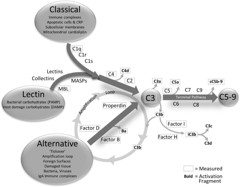
Purpose: Age-related macular degeneration (AMD) is an acquired degenerative disease of the retina classified into early, intermediate, and advanced AMD. A key factor in the pathogenesis of AMD is the complement system. The interaction of age and sex with the complement system may affect the risk of developing AMD. The purpose of this study was to determine if there were sex-specific differences in levels of complement factors among patients with the intermediate phenotype of AMD (iAMD) and explore the correlation between age and complement proteins.

Methods: We studied complement factors in patients with iAMD and controls without AMD. Nonparametric, rank-based linear regressions including a sex by AMD interaction were used to compare levels for each analyte. Correlations between age and complement proteins were evaluated using the Spearman rank correlation coefficient.

Results: We found significantly higher levels of factor B and factor I in females compared with males with iAMD, whereas no differences were seen in complement levels in male and female controls. The ratios of Ba/factor B, C3a/C3, C4b/C4, and C5a/C5 were not different in males and females with iAMD.

Conclusions: We demonstrate disparities in a subset of systemic complement factors between females and males with iAMD, but apparent complement turnover as measured by ratios of activation fragments to intact molecules was not different between these groups. The results suggest that complement system levels, including complement regulator factor I, exhibits sex-related differences in patients with iAMD and highlights that stratification by sex might be helpful in the interpretation of clinical trials of anticomplement therapy.


Conflict of interest statement

Disclosure: A.I. Marin, None; F. Poppelaars, None; B.D. Wagner, None; A.G. Palestine, None; J.L. Patnaik, None; V.M. Holers, None; A.A. Frazer-Abel, None; M.T. Mathias, None; N. Manoharan, None; C.N. Fonteh, None; N. Mandava, None; A.M. Lynch, None

Figures

Figure 1.
Figure 1.
The classic, lectin, and alternative complement pathways. These three pathways are activated differently but all connect at the central point C3. The three pathways are interrelated by the action of AP as an amplification loop for the classical and lectin pathways. After C3, the complement system enters the terminal pathway, culminating in the membrane attached complex (also known as C5b-9). As activation flows through the cascade, the components are cleaved yielding activation fragments that are released into circulation. Note. Reprinted with permission from BMJ Open Ophth. Lynch AM, et al 2020;5:e000361 page 2. License date Nov 13,2021.
Figure 2.
Figure 2.
Boxplot showing difference among groups in complement components found to be significantly higher in females compared with males in patients with intermediate AMD. The boxplots extend to the 25th and 75th percentiles. The line represents the median and the large black pluses correspond to the mean values. Individual colored circles illustrate the raw values. Male patients are represented as blue circles and female patients are represented in red circles. “ns” represents no statistical difference between groups. All comparisons are adjusted for batch effects.

Similar articles

Cited by

References

    1. Wong WL, Su X, Li X, et al. .. Global prevalence of age-related macular degeneration and disease burden projection for 2020 and 2040: a systematic review and meta-analysis. Lancet Glob Health. 2014; 2(2): e106–e116, doi:10.1016/S2214-109X(13)70145-1. - DOI - PubMed
    1. Ferris FL 3rd, Wilkinson CP, Bird A, et al. .. Clinical classification of age-related macular degeneration. Ophthalmology. 2013; 120(4): 844–851, doi:10.1016/j.ophtha.2012.10.036. - DOI - PMC - PubMed
    1. Lambert NG, ElShelmani H, Singh MK, et al. .. Risk factors and biomarkers of age-related macular degeneration. Prog Retin Eye Res. 2016; 54: 64–102, doi:10.1016/j.preteyeres.2016.04.003. - DOI - PMC - PubMed
    1. Pennington KL, DeAngelis MM.. Epidemiology of age-related macular degeneration (AMD): associations with cardiovascular disease phenotypes and lipid factors. Eye Vis (Lond). 2016; 3: 34, doi:10.1186/s40662-016-0063-5. - DOI - PMC - PubMed
    1. Weber BH, Charbel Issa P, Pauly D, Herrmann P, Grassmann F, Holz FG. The role of the complement system in age-related macular degeneration. Dtsch Arztebl Int. 2014; 111(8): 133–138, doi:10.3238/arztebl.2014.0133. - DOI - PMC - PubMed

Publication types