. 2022 May 24;119(21):e2123208119.
doi: 10.1073/pnas.2123208119. Epub 2022 May 20.

MERS-CoV endoribonuclease and accessory proteins jointly evade host innate immunity during infection of lung and nasal epithelial cells

Affiliations

MERS-CoV endoribonuclease and accessory proteins jointly evade host innate immunity during infection of lung and nasal epithelial cells

Courtney E Comar et al. Proc Natl Acad Sci U S A. .

Abstract

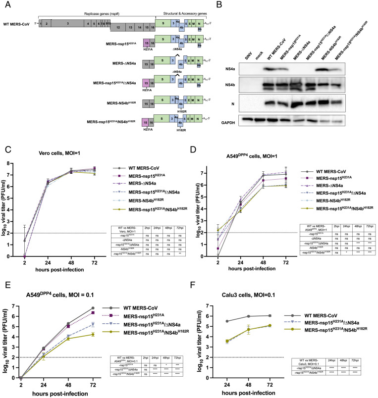
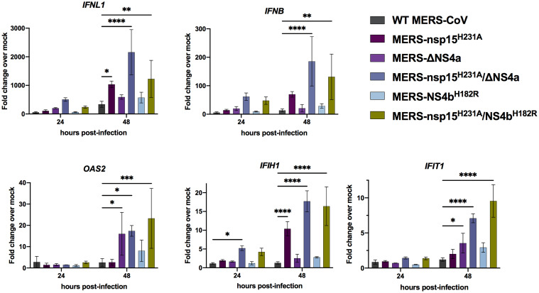
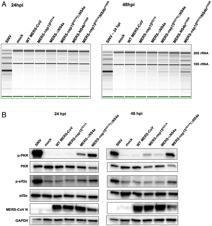
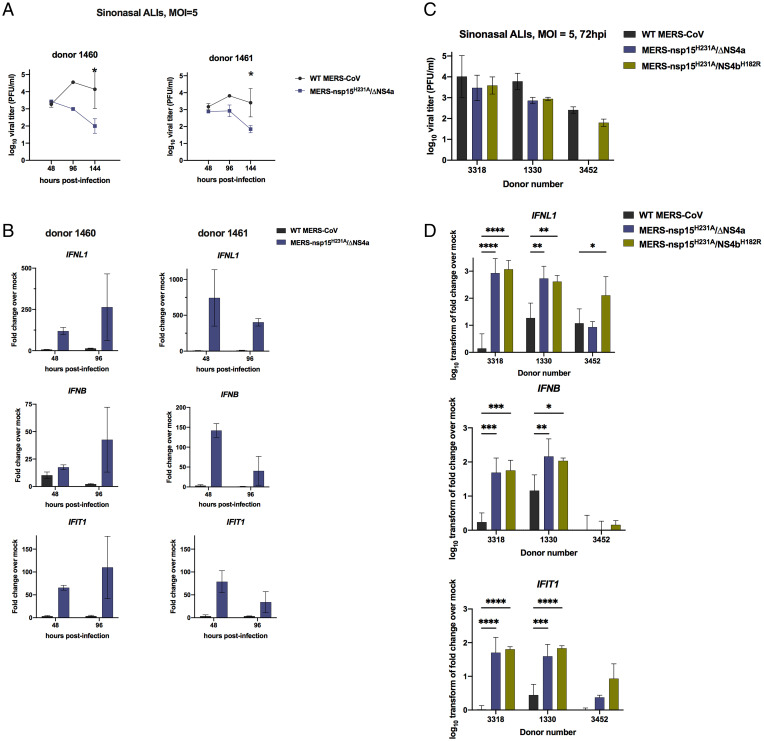
Middle East respiratory syndrome coronavirus (MERS-CoV) emerged into humans in 2012, causing highly lethal respiratory disease. The severity of disease may be, in part, because MERS-CoV is adept at antagonizing early innate immune pathways—interferon (IFN) production and signaling, protein kinase R (PKR), and oligoadenylate synthetase/ribonuclease L (OAS/RNase L)—activated in response to viral double-stranded RNA (dsRNA) generated during genome replication. This is in contrast to severe acute respiratory syndrome CoV-2 (SARS-CoV-2), which we recently reported to activate PKR and RNase L and, to some extent, IFN signaling. We previously found that MERS-CoV accessory proteins NS4a (dsRNA binding protein) and NS4b (phosphodiesterase) could weakly suppress these pathways, but ablation of each had minimal effect on virus replication. Here we investigated the antagonist effects of the conserved coronavirus endoribonuclease (EndoU), in combination with NS4a or NS4b. Inactivation of EndoU catalytic activity alone in a recombinant MERS-CoV caused little if any effect on activation of the innate immune pathways during infection. However, infection with recombinant viruses containing combined mutations with inactivation of EndoU and deletion of NS4a or inactivation of the NS4b phosphodiesterase promoted robust activation of dsRNA-induced innate immune pathways. This resulted in at least tenfold attenuation of replication in human lung–derived A549 and primary nasal cells. Furthermore, replication of these recombinant viruses could be rescued to the level of wild-type MERS-CoV by knockout of host immune mediators MAVS, PKR, or RNase L. Thus, EndoU and accessory proteins NS4a and NS4b together suppress dsRNA-induced innate immunity during MERS-CoV infection in order to optimize viral replication.

Keywords: MERS-CoV; PKR; RNase L; endonuclease U; innate immune antagonism.


Conflict of interest statement

Competing interest statement: S.R.W. is on the Scientific Advisory Boards of Immunome, Inc and Ocugen, Inc. N.A.C. consults for GSK, AstraZeneca, Novartis, Sanofi/Regeneron, and Oyster Point Pharmaceuticals and has US patent “Therapy and Diagnostics for Respiratory Infection” (10,881,698 B2, WO20913112865) and a licensing agreement with GeneOne Life Sciences. A.R.F. consults with Deciphera Inc.

Figures

Fig. 1.
Fig. 1.
Recombinant MERS-CoV design and replication kinetics. (A) Diagram of the MERS-CoV genome including the replicase locus (encoding 16 nsps), structural genes, and accessory genes is shown along with a diagram of each recombinant mutant constructed using the BAC reverse genetics system. (B) A549DPP4 cells were infected at MOI = 5 with the indicated viruses, and protein lysates were harvested at 24 hpi. Expression of NS4a and NS4b was determined by SDS/PAGE and Western blot. (C) Vero cells were infected in triplicate at MOI = 1 with the indicated viruses. Supernatants were collected at the indicated hpi, and infectious virus was quantified by plaque assay. (D) A549DPP4 cells were infected in triplicate with the indicated viruses at MOI = 1, and replication was quantified as in C. Data in C and D are from one representative of three independent experiments. (E) A549DPP4 and (F) Calu-3 cells were infected in triplicate with the indicated viruses at MOI = 0.1, and replication was quantified as in C. All data in CF are displayed as means ± SD. The dotted line in CF indicates the limit of detection of the plaque assay. Statistical significance of each recombinant virus compared to WT MERS-CoV was calculated by repeated measures two-way ANOVA: *P ≤ 0.05; **P ≤ 0.01; ***P ≤ 0.001; ****P ≤ 0.0001. Data that were not statistically significant are labeled ns.
Fig. 2.
Fig. 2.
The dsRNA expression is increased when EndoU is inactive during MERS-CoV infection. (A) Representative dsRNA staining in MERS-CoV infection: A549DPP4 cells were infected at MOI = 5 and fixed at 24 and 48 hpi with 4% paraformaldehyde and subjected to IF to detect dsRNA by 20× widefield microscopy. Nuclei were stained by Hoechst, shown in blue, and dsRNA was stained by J2, shown in white. (Scale bar, 50 μm.) (B) Representative images for quantification of dsRNA in A549DPP4 cells, mock infected (24 hpi), or MERS-CoV infected at MOI = 5 and fixed 48 hpi are shown with all single channels (dsRNA, nsp8, N+ssRNA, DAPI). Merged image shows DAPI (blue), nsp8 (magenta), and dsRNA (green). (Scale bar, 50 μm.) The dsRNA and nsp8 were stained by IF using J2 and anti-nsp8 serum, N+ssRNA was stained by FISH, and nuclei were stained by Hoechst (DAPI). (C) The dsRNA was quantified by graphing the ratio of dsRNA MGV over nsp8 MGV in each infected ROI. An ROI was defined by thresholding using the Otsu method for N+ssRNA staining in Fiji. Each dot represents the ratio of dsRNA/nsp8 for that ROI. Five to seven representative fields of widefield microscopy at 20× with 1.5× zoom were analyzed per condition. Data shown are from one representative of two independent experiments, and mean plus SD is shown. Statistical significance compared to WT MERS-CoV was determined by one-way ANOVA. *P ≤ 0.05; ****P ≤ 0.0001. Data that were not statistically significant are not labeled.
Fig. 3.
Fig. 3.
MERS-nsp15H231A/ΔNS4a and MERS-nsp15H231A/NS4bH182R induce IFN/ISG expression in A549DPP4 cells. A549DPP4 cells were mock infected or infected in triplicate at MOI = 5. Total RNA was harvested at 24 and 48 hpi, and expression of IFNL1, IFNB, IFIH1, OAS2, and IFIT1 mRNAs was quantified by qRT-PCR and expressed as fold change over mock infected using the 2−Δ(ΔCT) formula. Data are displayed as means ± SD. Data are from one representative of three independent experiments. Statistical significance compared to WT MERS-CoV was calculated by two-way ANOVA: *P ≤ 0.05; **P ≤ 0.01; ***P ≤ 0.001; ****P ≤ 0.0001. Data that were not statistically significant are not labeled.
Fig. 4.
Fig. 4.
RNase L and PKR are activated in the absence of EndoU activity combined with loss of accessory proteins during MERS-CoV infection. (A) A549DPP4 cells were mock infected or infected at MOI = 5, and total cellular RNA was harvested at 24 and 48 hpi. The rRNA degradation was assessed on an Agilent Bioanalyzer. 28S and 18S rRNA positions are indicated. Data are from one representative of three independent experiments. (B) A549DPP4 cells were mock infected or infected at MOI = 5 and cell lysates were harvested at 24 and 48 hpi. Proteins were separated by SDS/PAGE and immunoblotted with antibodies against phosphorylated PKR (p-PKR), PKR, phosphorylated eIF2α (p-eIF2α), eIF2α, MERS-CoV nucleocapsid (N), and glyceraldehyde-3-phosphate dehydrogenase (GAPDH). Data are from one representative of four (24 hpi) or two (48 hpi) independent experiments.
Fig. 5.
Fig. 5.
KO of innate immune pathways rescues attenuation in MERS-nsp15H231A/ΔNS4a and MERS-nsp15H231A/NS4bH182R. (A) Cell lysates of WT A549DPP4 as well as MAVS KO, PKR KO, and RNase L KO A549DPP4 were collected, and proteins were separated by SDS/PAGE and immunoblotted with antibodies against MAVS, PKR, RNase L, and GADPH. (B) WT A549DPP4 and MAVS KO A549DPP4 cells were infected with Sendai virus at MOI = 5, and total cellular RNA was harvested at 18 hpi. Expression of IFNL1 and IFNB was quantified by qRT-PCR, and CT values were normalized to beta-actin and shown as fold change over mock using the formula 2-Δ(ΔCT). Data are displayed as means ± SD. (C) A549DPP4 cell lines: WT, MAVS KO, PKR KO, and RNase L KO were infected in triplicate at MOI = 1, and supernatant samples were collected at 48 hpi. Viral replication was quantified by plaque assay. Data are displayed as means ± SD. Statistical significance of differences in viral replication for each recombinant virus compared to WT MERS-CoV was calculated by repeated measures two-way ANOVA: *P ≤ 0.05; **P ≤ 0.01; ****P ≤ 0.0001. Data that were not statistically significant are not labeled. Data are from one representative of three independent experiments.
Fig. 6.
Fig. 6.
MERS-nsp15H231A/ΔNS4a and MERS-nsp15H231A/NS4bH182R are attenuated and induce IFN/ISGs in primary nasal epithelial cells. (A) Nasal air–liquid interface (ALI) cultures derived from two independent donors were infected in duplicate on the apical cell surface at MOI = 5. Apically released virus was collected at 48, 96, and 144 hpi and quantified by plaque assay. (B) After apically released virus was collected from each transwell at the indicated time, total cellular RNA was collected, and IFN and ISG mRNA expression was quantified via qRT-PCR. CT values were normalized to 18S rRNA and shown as fold change over mock using the formula 2-Δ(ΔCT). (C) Nasal ALI cultures from three independent donors were infected apically in triplicate at MOI = 5, and apically released virus was collected at 72 hpi for quantification by viral plaque assay. (D) After collecting apically shed virus, total cellular RNA was collected, and IFN and IFIT1 mRNA expression was quantified via qRT-PCR as detailed above using the 2-Δ(ΔCT) method. Data were then log10 transformed and displayed as mean ± SD. Statistical significance of differences were calculated by repeated measures two-way ANOVA: *P ≤ 0.05; **P ≤ 0.01; ***P ≤ 0.001; ****P ≤ 0.0001. Data that were not statistically significant are not labeled.

Update of

References

    1. Zaki A. M., van Boheemen S., Bestebroer T. M., Osterhaus A. D., Fouchier R. A., Isolation of a novel coronavirus from a man with pneumonia in Saudi Arabia. N. Engl. J. Med. 367, 1814–1820 (2012). - PubMed
    1. Khalafalla A. I., et al. , MERS-CoV in upper respiratory tract and lungs of dromedary camels, Saudi Arabia, 2013-2014. Emerg. Infect. Dis. 21, 1153–1158 (2015). - PMC - PubMed
    1. Reusken C. B. E. M., et al. , Geographic distribution of MERS coronavirus among dromedary camels, Africa. Emerg. Infect. Dis. 20, 1370–1374 (2014). - PMC - PubMed
    1. Wernery U., et al. , A phylogenetically distinct Middle East respiratory syndrome coronavirus detected in a dromedary calf from a closed dairy herd in Dubai with rising seroprevalence with age. Emerg. Microbes Infect. 4, e74 (2015). - PMC - PubMed
    1. Corman V. M., et al. , Rooting the phylogenetic tree of Middle East respiratory syndrome coronavirus by characterization of a conspecific virus from an African bat. J. Virol. 88, 11297–11303 (2014). - PMC - PubMed

Publication types

MeSH terms

Substances