Review
. 2022 Jun;17(6):890-901.
doi: 10.2215/CJN.13341021. Epub 2022 May 20.

Mechanical Circulatory Support: Primer for Consultant Specialists

Affiliations
Review

Mechanical Circulatory Support: Primer for Consultant Specialists

Christopher W Tam et al. Clin J Am Soc Nephrol. 2022 Jun.

Abstract

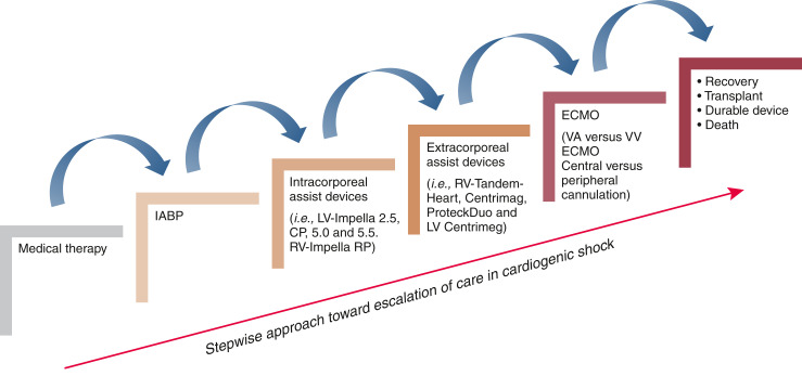
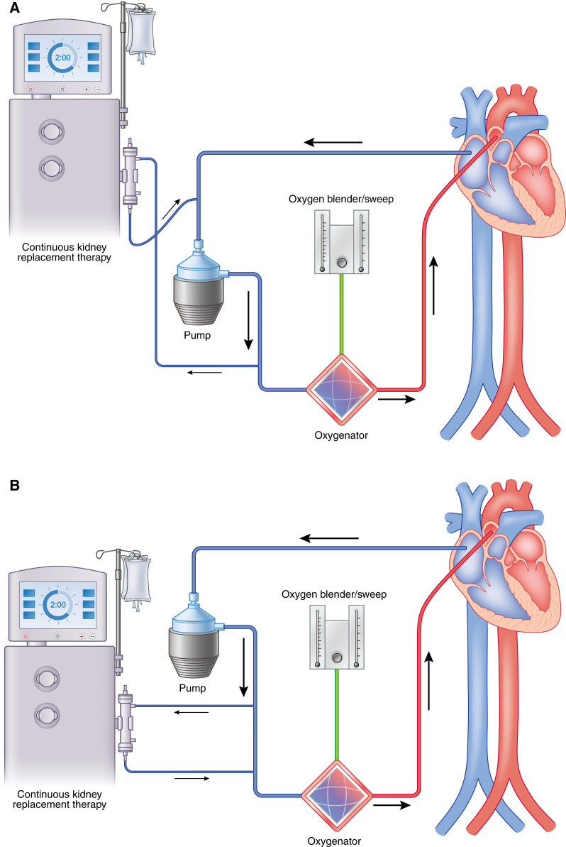
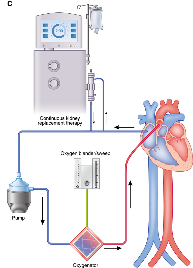
Mechanical life support therapies exist in many forms to temporarily replace the function of vital organs. Generally speaking, these tools are supportive therapy to allow for organ recovery but, at times, require transition to long-term mechanical support. This review will examine nonrenal extracorporeal life support for cardiac and pulmonary support as well as other mechanical circulatory support options. This is intended as a general primer and overview to assist nephrologist consultants participating in the care of these critically ill patients who often experience acute renal injury as a result of cardiopulmonary shock and from their exposure to mechanical circulatory support.

Keywords: cardiovascular; cardiovascular disease; critical care nephrology and acute kidney injury series; heart failure.


Figures

Figure 1.
Figure 1.
Stepwise escalation of care in cardiogenic shock. ECMO, extracorporeal membrane oxygenation; IABP, intra-aortic balloon pump; LV, left ventricular; RV, right ventricular; VA ECMO, venoarterial extracorporeal membrane oxygenation; VV ECMO, venovenous extracorporeal membrane oxygenation.
Figure 2.
Figure 2.
Diagram of an Impella 5.5 for LV support. Adapted from Abiomed, Inc., Danvers, MA, with permission.
Figure 3.
Figure 3.
Diagram of an Impella RP for RV support. Adapted from Abiomed, Inc., Danvers, MA, with permission.
Figure 4.
Figure 4.
Syncardia total artificial heart (TAH) device. The left panel represents a depiction of postplacement TAH with anastomosis to both the right and left atria as well as the main pulmonary artery and aorta. The right panel represents a depiction of a human heart preimplantation of TAH. Adapted from ref. , with permission.
Figure 5.
Figure 5.
ECMO may be configured either peripherally or centrally with different risks and benefits of either choice. (A) Diagram demonstrating central cannulation for VA ECMO. Right atrium/Inferior Vena Cava (IVC) is cannulated, and blood is flowing to the ECMO pump into the oxygenator and flowing back into the patient in an antegrade fashion directly into the ascending aorta. (B) Diagram demonstrating peripheral cannulation for VA ECMO. The femoral vein is cannulated, and blood is flowing from the patient to the ECMO pump into the oxygenator and flowing back into the patient in a retrograde fashion via the femoral artery. For (A) and (B), the oxygen blender and sweep as shown can be titrated accordingly on the basis of the partial pressure of oxygen in arterial blood (PaO2) and the partial pressure of carbon dioxide in arterial blood (PaCO2). This is the most invasive type of cannulation for ECMO. (C) Diagram demonstrating North-South syndrome or Harlequinn syndrome as seen in patients with peripheral VA ECMO. The mixing cloud occurs at the junction of retrograde oxygenation blood from the ECMO with that of the patient’s native LV cardiac output. Blood proximal to the mixing cloud is often deoxygenated, hence the importance of a right upper extremity arterial line in peripheral VA ECMO. (D) Diagram demonstrating VV ECMO with a right internal jugular vein and right femoral vein cannulation. Blood is flowing from the right femoral vein to the ECMO pump into the oxygenator and returning back into the patient in the right internal jugular vein.
Figure 6.
Figure 6.
Continuous kidney replacement therapy may be incorporated into the ECMO circuit in place of using a dialysis catheter. (A) Continuous KRT (CKRT) machine with venous limb connected post-ECMO pump and the arterial limb in the pre-ECMO pump but before the oxygenator. (B) The CKRT machine with venous and arterial limb post-ECMO pump but before the oxygenator. (C) The CKRT machine with venous and arterial lines pre-ECMO pump and before the oxygenator. It is important to maintain the CKRT circuit, especially the return limb before the oxygenator, to prevent air embolization, venous admixture, and pressure elevations in the ECMO circuit (58).
Figure 6.
Figure 6.
Continuous kidney replacement therapy may be incorporated into the ECMO circuit in place of using a dialysis catheter. (A) Continuous KRT (CKRT) machine with venous limb connected post-ECMO pump and the arterial limb in the pre-ECMO pump but before the oxygenator. (B) The CKRT machine with venous and arterial limb post-ECMO pump but before the oxygenator. (C) The CKRT machine with venous and arterial lines pre-ECMO pump and before the oxygenator. It is important to maintain the CKRT circuit, especially the return limb before the oxygenator, to prevent air embolization, venous admixture, and pressure elevations in the ECMO circuit (58).

References

    1. Thiele H, Zeymer U, Neumann FJ, Ferenc M, Olbrich HG, Hausleiter J, Richardt G, Hennersdorf M, Empen K, Fuernau G, Desch S, Eitel I, Hambrecht R, Fuhrmann J, Böhm M, Ebelt H, Schneider S, Schuler G, Werdan K; IABP-SHOCK II Trial Investigators : Intraaortic balloon support for myocardial infarction with cardiogenic shock. N Engl J Med 367: 1287–1296, 2012 - PubMed
    1. Hochman JS, Sleeper LA, Webb JG, Sanborn TA, White HD, Talley JD, Buller CE, Jacobs AK, Slater JN, Col J, McKinlay SM, LeJemtel TH: Early revascularization in acute myocardial infarction complicated by cardiogenic shock. N Engl J Med 341: 625–634, 1999 - PubMed
    1. Ponikowski P, Voors AA, Anker SD, Bueno H, Cleland JGF, Coats AJS, Falk V, González-Juanatey JR, Harjola VP, Jankowska EA, Jessup M, Linde C, Nihoyannopoulos P, Parissis JT, Pieske B, Riley JP, Rosano GMC, Ruilope LM, Ruschitzka F, Rutten FH, van der Meer P; ESC Scientific Document Group : 2016 ESC Guidelines for the diagnosis and treatment of acute and chronic heart failure [published correction appears in Eur Heart J 2016;38:ehw383]. Eur Heart J 37: 2129–2200, 2016 - PubMed
    1. McMurray JJ, Pfeffer MA: Heart failure. Lancet 365: 1877–1889, 2005 - PubMed
    1. Tanai E, Frantz S: Pathophysiology of heart failure. Compr Physiol 6: 187–214, 2015 - PubMed