. 2022 May 20;8(1):47.
doi: 10.1038/s41421-022-00405-2.

Structural insights into the activation of somatostatin receptor 2 by cyclic SST analogues

Affiliations

Structural insights into the activation of somatostatin receptor 2 by cyclic SST analogues

Qing Bo et al. Cell Discov. .

Abstract

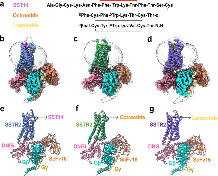
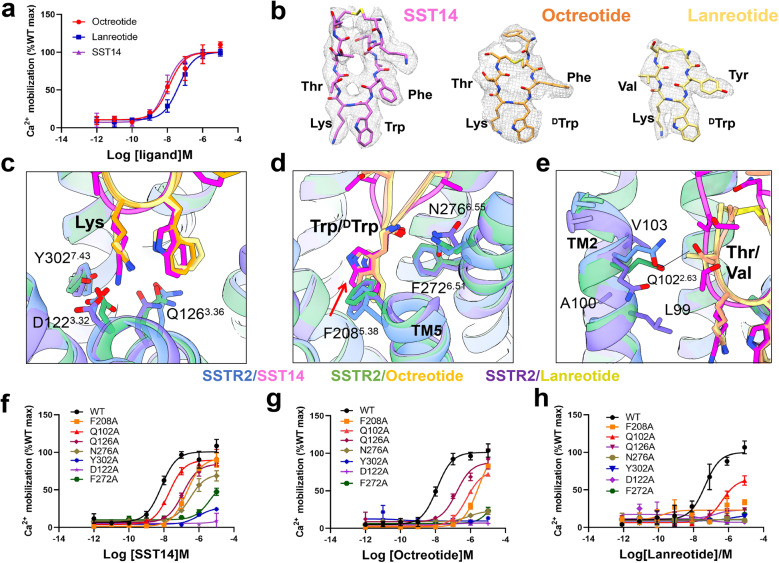
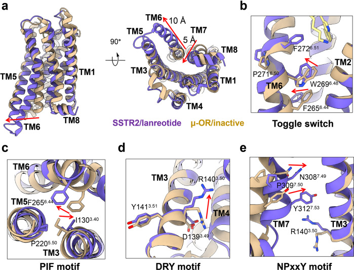
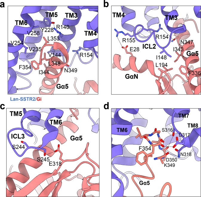
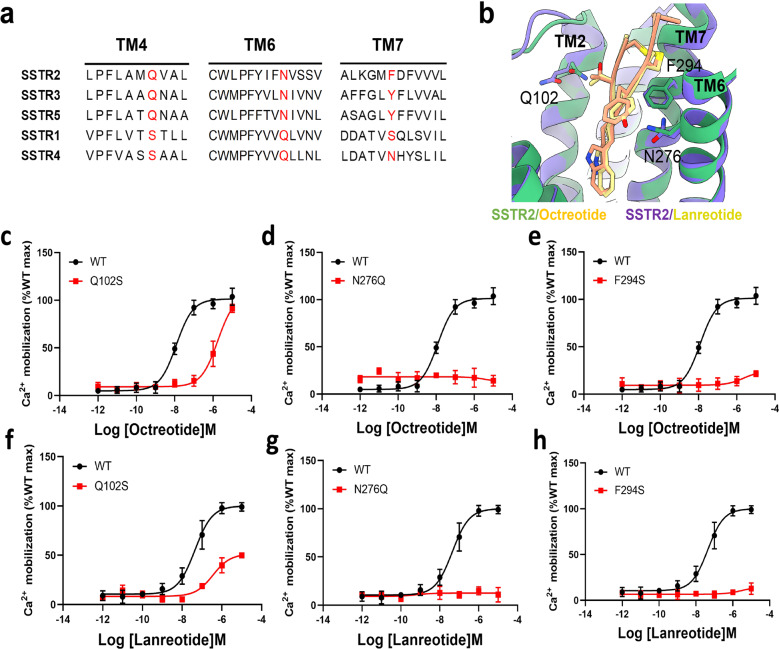
The endogenous cyclic tetradecapeptide SST14 was reported to stimulate all five somatostatin receptors (SSTR1-5) for hormone release, neurotransmission, cell growth arrest and cancer suppression. Two SST14-derived short cyclic SST analogues (lanreotide or octreotide) with improved stability and longer lifetime were developed as drugs to preferentially activate SSTR2 and treat acromegalia and neuroendocrine tumors. Here, cryo-EM structures of the human SSTR2-Gi complex bound with SST14, octreotide or lanreotide were determined at resolutions of 2.85 Å, 2.97 Å, and 2.87 Å, respectively. Structural and functional analysis revealed that interactions between β-turn residues in SST analogues and transmembrane SSTR2 residues in the ligand-binding pocket are crucial for receptor binding and functional stimulation of the two SST14-derived cyclic octapeptides. Additionally, Q1022.63, N2766.55, and F2947.35 could be responsible for the selectivity of lanreotide or octreotide for SSTR2 over SSTR1 or SSTR4. These results provide valuable insights into further rational development of SST analogue drugs targeting SSTR2.


Conflict of interest statement

The authors declare no competing interests.

Figures

Fig. 1
Fig. 1. The cryo-EM structure of SSTR2–DNGi complex.
a Peptide sequence alignment of SST14, octreotide and lanreotide. bd Cryo-EM density maps of SST14 (b) or octreotide (c) or lanreotide (d) bound SSTR2–DNGi complex. eg Ribbon diagram representation of the cryo-EM structures of SST14 (e) or octreotide (f) or lanreotide (g) bound SSTR2–DNGi complex, colored by subunit (TMDs in sea green, SST14 in magenta, octreotide in orange, lanreotide in yellow, Gαi in hot pink, Gβ in cyan, Gγ in gold, ScFv16 in salmon).
Fig. 2
Fig. 2. Interactions between SST β-turn residues and SSTR2 transmembrane residues.
a Ca2+ response of SSTR2 with SST14, octreotide, and lanreotide. Data are presented as means ± SEM of three independent experiments conducted in triplicate. b Local density maps of SST14, octreotide and lanreotide. The conserved key binding amino acids are highlighted. ce Superposition of the ligand-binding pockets of SST14- or octreotide- or lanreotide-bound SSTR2 receptor. fh Ca2+ accumulation analysis of wild-type (WT) SSTR2 and mutants with SST14 (f) or octreotide (g) or lanreotide (h). Site mutations around the ligand-binding pocket disrupted the receptor–ligand interactions, resulting in SSTR2 malfunction in the Ca2+ accumulation assay. Data are presented as means ± SEM of three independent experiments conducted in triplicate.
Fig. 3
Fig. 3. Conformational change analysis of SSTR2 receptor upon binding of lanreotide.
a Comparision of lanreotide-bound SSTR2 (purple) and inactive state μ-OR (tan). be Conformational changes of toggle switch (b), PIF motif (c), DRY motif (d) and NPxxY motif (e) after SSTR2 activation upon binding of lanreotide.
Fig. 4
Fig. 4. Interaction interfaces between the SSTR2 and Gi protein in the presence of lanreotide.
a The network of interactions between lanreotide-bound SSTR2 (purple) and α5 helix of Gi (salmon). b The ICL2–Gi interface of lanreotide-bound SSTR2 (purple) and Gi (salmon). c The ICL3–Gi interface of lanreotide-bound SSTR2 (purple) and Gi (salmon). d The TM7–TM8 loop and α5 helix interface of lanreotide-bound SSTR2 (purple) and Gi (salmon).
Fig. 5
Fig. 5. Structural and functional presentation of subtype-selective site of SSTR2.
a Local sequence alignment of TM2, TM6, and TM7 of SSTR family. b Amino acid sites that may be associated with SSTR2 subtype selectivity are shown in the binding pockets of octreotide and lanreotide. ce Ca2+ accumulation analysis of wild-type (WT) SSTR2 and mutants with octreotide. Site mutations to the corresponding amino acids of SSTR1 disrupted the receptor–ligand interactions, resulting in SSTR2 malfunction in the Ca2+ accumulation assay. Data are presented as means ± SEM of three independent experiments conducted in triplicate. fh Ca2+ accumulation analysis of WT SSTR2 and mutants with lanreotide. Mutations to the corresponding amino acids of SSTR1 disrupted the receptor–ligand interactions, resulting in SSTR2 malfunction in the Ca2+ accumulation assay. Data are presented as means ± SEM of three independent experiments conducted in triplicate.

References

    1. Theodoropoulou M, Stalla GK. Somatostatin receptors: from signaling to clinical practice. Front. Neuroendocrinol. 2013;34:228–252. doi: 10.1016/j.yfrne.2013.07.005. - DOI - PubMed
    1. Møller LN, Stidsen CE, Hartmann B, Holst JJ. Somatostatin receptors. Biochimica et. Biophysica Acta. 2003;1616:1–84. doi: 10.1016/S0005-2736(03)00235-9. - DOI - PubMed
    1. Rocheville M, et al. Subtypes of the somatostatin receptor assemble as functional homo- and heterodimers. J. Biol. Chem. 2000;275:7862–7869. doi: 10.1074/jbc.275.11.7862. - DOI - PubMed
    1. Cuevas-Ramos D, Fleseriu M. Somatostatin receptor ligands and resistance to treatment in pituitary adenomas. J. Mol. Endocrinol. 2014;52:R223–R240. doi: 10.1530/JME-14-0011. - DOI - PubMed
    1. Watt HL, Kharmate G, Kumar U. Biology of somatostatin in breast cancer. Mol. Cell Endocrinol. 2008;286:251–261. doi: 10.1016/j.mce.2008.01.006. - DOI - PubMed