. 2022 May 20;12(1):8580.
doi: 10.1038/s41598-022-12206-4.

D-galactose-induced aging aggravates obesity-induced bone dyshomeostasis

Affiliations

D-galactose-induced aging aggravates obesity-induced bone dyshomeostasis

Napatsorn Imerb et al. Sci Rep. .

Abstract

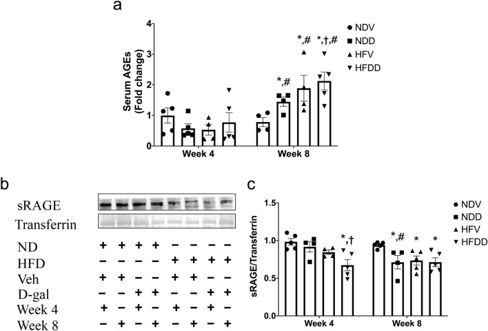
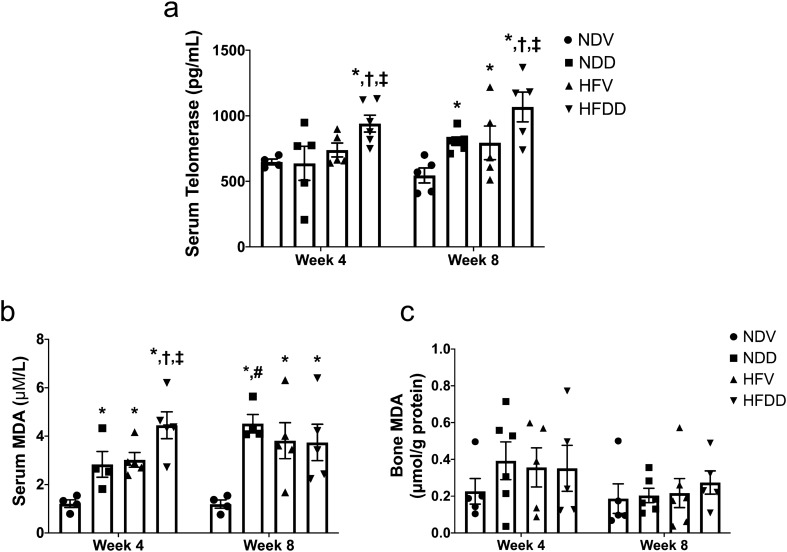
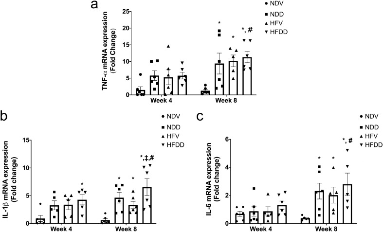
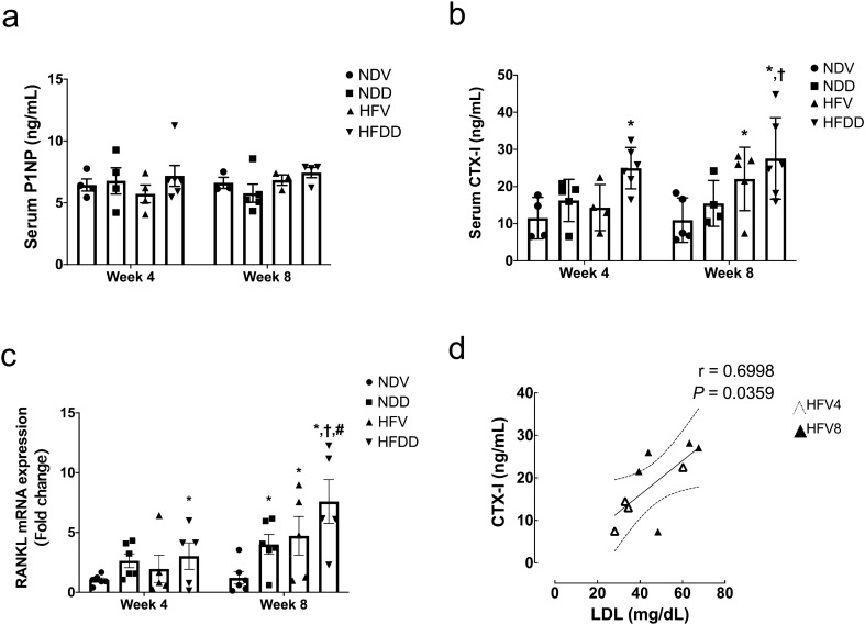
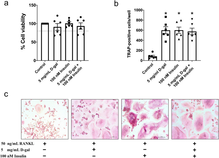
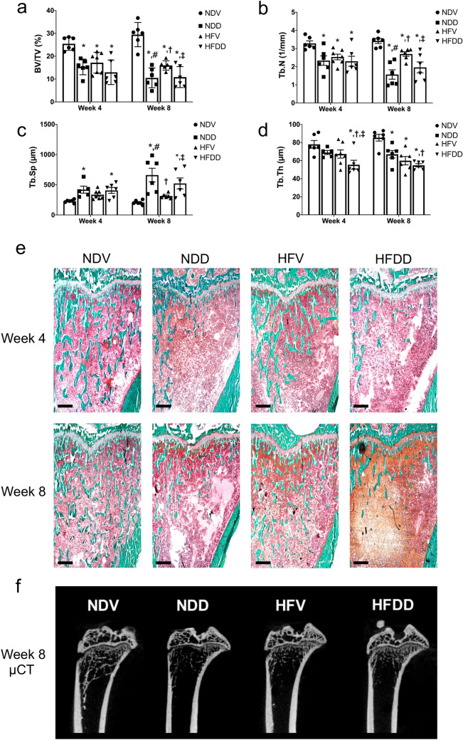
We aimed to compare the time-course effect of D-galactose (D-gal)-induced aging, obesity, and their combined effects on bone homeostasis. Male Wistar rats were fed with either a normal diet (ND; n = 24) or a high-fat diet (HFD; n = 24) for 12 weeks. All rats were then injected with either vehicle or 150 mg/kg/day of D-gal for 4 or 8 weeks. Blood was collected to measure metabolic, aging, oxidative stress, and bone turnover parameters. Bone oxidative stress and inflammatory markers, as well as bone histomorphometry were also evaluated. Additionally, RAW 264.7 cells were incubated with either D-gal, insulin, or D-gal plus insulin to identify osteoclast differentiation capacity under the stimulation of receptor activator of nuclear factor κB ligand. At week 4, D-gal-induced aging significantly elevated serum malondialdehyde level and decreased trabecular thickness in ND- and HFD-fed rats, when compared to the control group. At week 8, D-gal-induced aging further elevated advanced glycation end products, increased bone inflammation and resorption, and significantly impaired bone microarchitecture in HFD-fed rats. The osteoclast number in vitro were increased in the D-gal, insulin, and combined groups to a similar extent. These findings suggest that aging aggravates bone dyshomeostasis in the obese condition in a time-dependent manner.


Conflict of interest statement

The authors declare no competing interests.

Figures

Figure 1
Figure 1
D-galactose-induced aging aggravated aging-related biomarkers in the obese-insulin resistant condition. (a) Quantification of serum AGEs level in NDV, NDD, HFV, HFDD at 4-week and 8-week timepoints, (b, c) sRAGE protein expression relative to transferrin as analyzed by western blotting. Bar graphs presented as mean ± SEM (n = 4–6/group). * p < 0.05 versus NDV at the same time point, † p < 0.05 versus NDD at the same time point, ‡ p < 0.05 versus HFV at the same time point, # p < 0.05 versus same group at the different time point. AGEs, advanced glycation end products; HFDD, high-fat diet with D-galactose; HFV, high-fat diet with vehicle; NDD, normal diet with D-galactose; NDV, normal diet with vehicle; sRAGE, soluble receptor for advanced glycation end products.
Figure 2
Figure 2
D-galactose-induced aging aggravated systemic oxidative stress in the obese-insulin resistant condition. (a) Levels of telomerase in serum at the indicated time points, (b) Levels of MDA concentration in serum at the indicated time points, (c) Levels of MDA concentration in skeletal tissue at the indicated time points. Bar graphs presented as mean ± SEM (n = 4–6/group). * p < 0.05 versus NDV at the same time point, † p < 0.05 versus NDD at the same time point, ‡ p < 0.05 versus HFV at the same time point, # p < 0.05 versus same group at the different time point. HFDD, high-fat diet with D-galactose; HFV, high-fat diet with vehicle; MDA, Malondialdehyde; NDD, normal diet with D-galactose; NDV, normal diet with vehicle.
Figure 3
Figure 3
D-galactose-induced aging aggravated bone inflammation in the obese-insulin resistant condition. (a) TNF-α mRNA expression in bone tissues at the indicated time points, (b) IL-1 β mRNA expression in bone tissues at the indicated time points, (c) IL-6 mRNA expression in bone tissues at the indicated time points. Bar graphs are presented as mean ± SEM (n = 5–6/group). * p < 0.05 versus NDV at the same time point, † p < 0.05 versus NDD at the same time point, ‡ p < 0.05 versus HFV at the same time point, # p < 0.05 versus same group at the different time point. HFDD, high-fat diet with D-galactose; HFV, high-fat diet with vehicle; IL-1 β, interleukin-1β; IL-6, interleukin-6; NDD, normal diet with D-galactose; NDV, normal diet with vehicle: TNF-α, Tumor necrosis factor alpha.
Figure 4
Figure 4
D-galactose-induced aging aggravated bone resorption markers in the obese-insulin resistant condition. (a) Levels of P1NP in serum at the indicated time points, (b) Levels of CTX-I in serum at the indicated time points. Bar graphs are presented as mean ± SEM (n = 4–6/ group). (c) RANKL mRNA expression in bone tissues at the indicated time points. (d) Correlation between CTX-I and LDL. * p < 0.05 versus NDV at the same time point, † p < 0.05 versus NDD at the same time point,p < 0.05 versus HFV at the same time point, # p < 0.05 versus same group at the different time point. CTX-I, C-terminal telopeptide of type I collagen; HFDD, high-fat diet with D-galactose; HFV, high-fat diet with vehicle; NDD, normal diet with D-galactose; NDV, normal diet with vehicle; P1NP, procollagen type I N-terminal propeptide; RANKL, receptor activator of nuclear factor κB ligand.
Figure 5
Figure 5
D-galactose-induced senescence and hyperinsulinism-stimulated insulin resistance accelerated RANKL-induced osteoclast differentiation in RAW 264.7 cells. (a) % cell viability of RAW 264.7 belonging to four treatment group, (b) Number of TRAP-positive multinucleated cells which presented more than three nuclei indicated a mature osteoclast. (c) TRAP positive staining of RANKL-stimulated multinucleated osteoclasts with the treatments of control, 5 mg/mL D-galactose, 100 nM insulin and the combined treatment (20 × magnification). Bar graphs presented as mean ± SEM (n = 6/group).* p < 0.05 versus control group. D-gal, D-galactose; RANKL, receptor activator of nuclear factor κB ligand; TRAP, tartrate-resistant acid phosphatase.
Figure 6
Figure 6
Synergistic effect of D-galactose-induced aging in obese-insulin resistance influences the impairment of bone microarchitecture and severe bone volume loss. (a) Quantification of trabecular bone volume per tissue volume (BV/TV) at the indicated time points, (b) Quantification of trabecular number (Tb.N) at the indicated time points, (c) Levels of trabecular separation (Tb.Sp) at the indicated time points, (d) Levels of trabecular thickness (Tb.Th) at the indicated time points, (e) Images of right tibiae of rats with Goldner’s trichrome staining histomorphometry belonging to eight groups at indicated time points (n = 6/group). (f) Representative images show epiphyseal and metaphyseal regions of tibiae from 8-week 0.9% NaCl or d-galactose-injected rats. The images were obtained from ultra-high-resolution microcomputed tomography (μCT). Scale bars = 1000 µM (× 20 magnification). Bar graphs presented as mean ± SEM. * p < 0.05 versus NDV at the same time point, † p < 0.05 versus NDD at the same time point, ‡ p < 0.05 versus HFV at the same time point, # p < 0.05 versus same group at the different time point. BV/TV, bone volume per tissue volume; HFDD, high-fat diet with D-galactose; HFV, high-fat diet with vehicle; NDD, normal diet with D-galactose; NDV, normal diet with vehicle; Tb.N, trabecular number; Tb.Sp, trabecular separation; Tb.Th, trabecular thickness.

References

    1. Johnell O, Kanis J. Epidemiology of osteoporotic fractures. Osteoporos. Int. 2005;16(Suppl 2):S3–7. doi: 10.1007/s00198-004-1702-6. - DOI - PubMed
    1. Pérez LM, et al. 'Adipaging': ageing and obesity share biological hallmarks related to a dysfunctional adipose tissue. J. Physiol. 2016;594:3187–3207. doi: 10.1113/JP271691. - DOI - PMC - PubMed
    1. Salvestrini V, Sell C, Lorenzini A. Obesity may accelerate the aging process. Front. Endocrinol. (Lausanne) 2019;10:266. doi: 10.3389/fendo.2019.00266. - DOI - PMC - PubMed
    1. Moser SC, van der Eerden BCJ. Osteocalcin—a versatile bone-derived hormone. Front. Endocrinol. (Lausanne) 2019;9:794. doi: 10.3389/fendo.2018.00794. - DOI - PMC - PubMed
    1. Patsch JM, et al. Increased bone resorption and impaired bone microarchitecture in short-term and extended high-fat diet–induced obesity. Metab Clin Exp. 2011;60:243–249. doi: 10.1016/j.metabol.2009.11.023. - DOI - PMC - PubMed

Publication types