. 2022 May 20;12(1):8515.
doi: 10.1038/s41598-022-12598-3.

ILDR1 promotes influenza A virus replication through binding to PLSCR1

Affiliations

ILDR1 promotes influenza A virus replication through binding to PLSCR1

Yueyue Liu et al. Sci Rep. .

Abstract

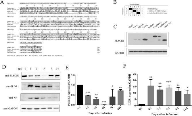
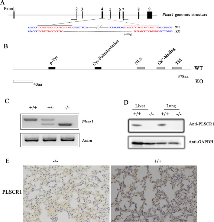
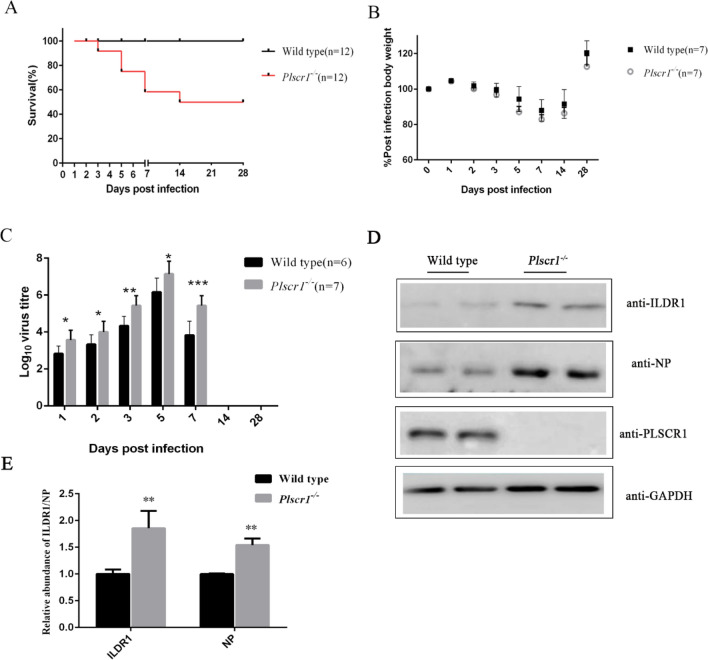
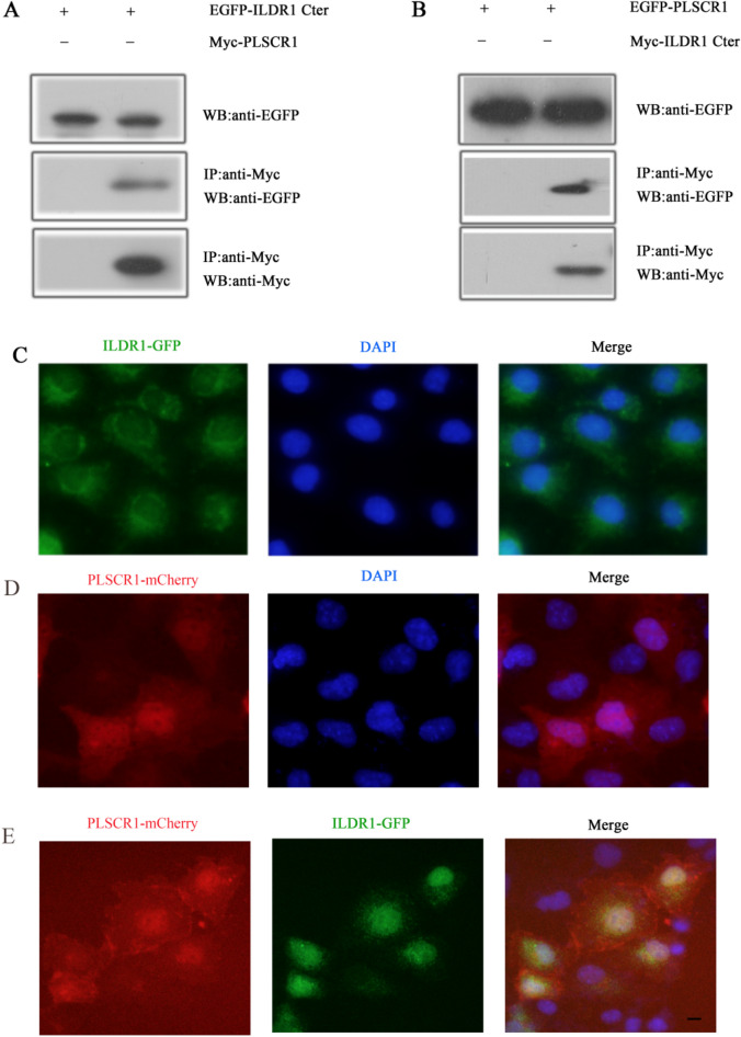
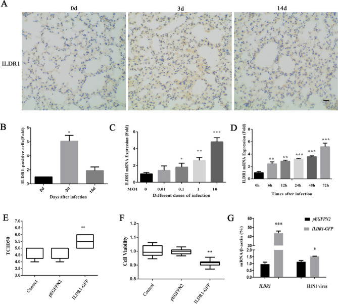
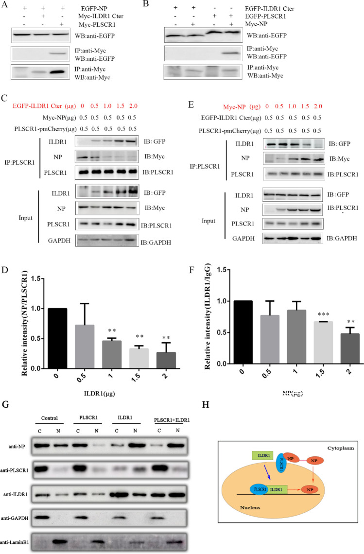
As a natural antiviral regulator, phospholipid scramblase 1 (PLSCR1) has been shown to inhibit influenza virus replication in infected cells through interacting with NP of influenza A virus (IAV). But its antiviral function as well as the underlying regulatory mechanism has not been examined in vivo. In the present work, we show that PLSCR1 expression is decreased in H1N1 SIV-infected mice, and Plscr1-/- mice are more susceptible to H1N1 SIV infection. By performing yeast two-hybrid screening, we identified immunoglobulin-like domain-containing receptor 1 (ILDR1) as a novel PLSCR1-binding partner. ILDR1 is highly expressed in the lungs, and its expression level is increased after virus infection. Interestingly, ILDR1 could not directly interact with virus NP protein, but could combine with PLSCR1 competitively. Our data indicates that there is a previously unidentified PLSCR1-ILDR1-NP regulatory pathway playing a vital role in limiting IAV infection, which provides novel insights into IAV-host interactions.


Conflict of interest statement

The authors declare no competing interests.

Figures

Figure 1
Figure 1
PLSCR1 Expression after H1N1 SIV Infection. (A) The similarity of PLSCR1 between different species. (B) The identity percentage of nucleoprotein of influenza virus between the isolated strain from the diseased pigs and human. (C) Western blots showed that PLSCR1 expression in various tissues of C57BL/6 mice on day 42. (D) PLSCR1 and ILDR1protein levels in lung are modulated after swine influenza A virus (SIV) infection. The lungs were dissected from C57BL6/J mice that were either mock- or SIV-infected at multiple days p.i. as indicated. (E and F) The area of the PLSCR1 and ILDR1 peak, in relation to the standard curve, was determined using ImageJ software (n = 3). Bars represent mean ± s.d. *P < 0.05, **P < 0.01; ***P < 0.001.
Figure 2
Figure 2
Construction of Plscr1 knockout mice. (A) The schematic drawing of the strategy for Plscr1 gene disruption. The target sites of clustered regularly interspaced short palindromic repeat (CRISPR)-Cas9 small guide RNAs (sgRNAs) in the Plscr1 gene are indicated in red, and the deleted region in the Plscr1 gene of knockout mice is indicated by dashes. The positions of RT-PCR primers are indicated by arrows. (B) The schematic drawing of the domain structure of PLSCR1 in wildtype and knockout mice. (C) The expression of Plscr1 mRNA in the tail of P42 mice was determined by RT-PCR. β-actin was included as the internal control. The corresponding size were respectively 636 bp, 514 bp corresponding to wild types and Plscr1-/- mice. (D) Western blot showed the expression in the liver and lung of P42 mice. (E) PLSCR1 was detected by immunohistology using rabbit anti-mPLSCR1, visualized with 3,3,-diaminobenzidine, and counterstained with hematoxylin. Scale bar: 20 μm. +/+ :wild type mice; +/− : heterozygous mice; −/−:Plscr1 knockout mice.
Figure 3
Figure 3
PLSCR1 influences the infection of mice by Swine influenza virus (SIV). (A) C57BL/6 J and Plscr1/ mice were infected intranasally with 103 pfu SIV. Suviral was determined daily and calculated as a percentage of the initial total numbers. (B) Mice were weighed daily and the weights represented as a percentage of the starting weight (n = 7 per group). (C) Lung tissues were taken at multiple days p.i. as indicated and virus titer determined by plaque assay (n > 6 per group). Data represent the mean value ± SD. Asterisks indicate statistical difference (Unpaired t test, *P < 0.05, **P < 0.01, ***P < 0.001). (D) Western blots showed that ILDR1 and NP protein expression in lung of C57BL/6 and Plscr1/ mice on day 42 after infected with 103 pfu SIV for 3 days. (E) Statistical analysis of ILDR1 and NP levels in Plscr1/ mice. The value for ILDR1 and NP were standardized to the GAPDH level and normalized to the level of ILDR1 and NP in lung of wild type mice. Data are shown as the means SD from three independent experiments (Unpaired t test, *P < 0.05, **P < 0.01, ***P < 0.001).
Figure 4
Figure 4
PLSCR1 binds ILDR1. (A) Western blots showing that EGFP-tagged cytoplasmic fragment of ILDR1 was co-immunoprecipitated with Myc-tagged PLSCR1. (B) Western blots showing that EGFP-taggedPLSCR1 was co-immunoprecipitated with Myc-tagged cytoplasmic fragment of ILDR1. Expression vectors were transfected into HEK293T cells to express epitope-tagged proteins, and cell lysis were subject to immunoprecipitation.5% of total protein was loaded as input. IP indicates antibody used for immunoprecipitation and WB indicates antibody used for detection. (C) ILDR1-EGFP localize in the cytoplasm of COS7. (D) PLSCR1-mCherry localize both in the nucleus and cytoplasm of COS7. (E) However, when PLSCR1-mCherry is present, ILDR1-GFP translocates into the nuclei. Expression vectors were transfected into COS-7 cells to express epitope-tagged proteins. Nuclei were stained with DAPI. Scale bar: 10 μm.
Figure 5
Figure 5
ILDR1 Expression after swine influenza virus infection. (A) C57BL/6 J mice were infected intranasally with 103 pfu. SIV. The lungs of mice were taken after infection 0, 3 and14 days for immunohistochemical detection. ILDR1 was detected by immunohistology using rabbit anti-ILDR1, visualized with 3,3,-diaminobenzidine and counterstained with hematoxylin. Scale bar: 20 μm. The expression levels of the ILDR1 after infection relative to that of normal expression were analyzed by areal density (B). (C) HEK 293 T cells were infected with different doses(MOI = 0,0.01,0.1,1,10) for 48 h, and RNA were extracted, then the expression of ILDR1 was confirmed by quantitative reverse-transcription PCR. Data represent the mean value ± SD. Asterisks indicate statistical difference (n = 3, Unpaired t test; *P < 0.05, **P < 0.01, ***P < 0.001). (D) Expression of ILDR1 in virus-infected cells at an MOI of 0.1. RNA were extracted at different time points (0 h, 6 h, 12 h, 24 h, 48 h) and subjected to RT-qPCR. Data represent the mean value ± SD. Asterisks indicate statistical difference (n = 3, Unpaired t test; *P < 0.05, **P < 0.01, ***P < 0.001). (E) Virus replication in ILDR1-overexpressing HEK293T cells. Cells were transfected with ILDR1-GFP or pEGFPN2 for 24 h, and then infected with SIV at an MOI of 0.1. RNA were extracted at the indicated time points, and virus titers or NP expression were determined by TCID50, CCK-8 (F) and RT-qPCR (G).
Figure 6
Figure 6
The viral PLSCR1-NP interaction is inhibited by ILDR1. (A, B) No interaction of ILDR1 with viral NP as determined by the co-IP assays. The interaction between the ILDR1 Cter domain and the NP of SIV was validated using the co-IP assays.HEK293T cells were transfected with ILDR1and NP protein. The Myc-tagged PLSCR1 plasmids were used as positive controls. All cell lysates were prepared at 24 h post transfection and proteins were immunoprecipitated using an anti-Myc mouse MAb, or anti-EGFP rabbit MAb. The immunoprecipitated proteins were analyzed by Western blotting. (C) HEK293T cells were transfected with Myc-NP (0.5 μg), PLSCR1-pmCherry (0.5 μg) and different concentrations of EGFP-ILDR1 cytoplasmic fragment (0 μg,0.5 μg,1 μg,1.5 μg,2 μg). The cell lysates were prepared, and proteins were immunoprecipitated using an anti-PLSCR1 rabbit PAb. The expression levels of the ILDR1 and NP protein after immunoprecipitation relative to that of GAPDH were analyzed by densitometry, n = 3 (D). (E) HEK293T cells were transfected with EGFP-ILDR1 cytoplasmic fragment (0.5 μg), PLSCR1-pmCherry (0.5 μg) and different concentrations of Myc-NP (0 μg,0.5 μg,1 μg,1.5 μg,2 μg). The cell lysates were prepared, and proteins were immunoprecipitated using an anti-PLSCR1 rabbit PAb. The expression levels of the ILDR1 and NP protein after immunoprecipitation relative to that of GAPDH were analyzed by densitometry, n = 3 (F). (G) Cells were transfected with ILDR1-GFP, PLSCR1-Myc or empty retrovirus-transfected control for 24 h, and then infected with SIV at an MOI of 0.1. At 6 h p.i., the cells were separated into nuclear (N) and cytoplasmic fractions (C). Each fraction was subjected to western blotting with corresponding antibody for protein detection. (H) Model of PLSCR1-NP interaction is competitively by ILDR1. ILDR1 located in the cytoplasm, when in the presence of PLSCR1, ILDR1 and PLSCR1 co-translocate into the nuclei. PLSCR1 could prevent the nuclear import of NP protein and ILDR1 could bind to PLSCR1 competitively with NP. So in the nuclei, the viral PLSCR1-NP interaction is inhibited by ILDR1.

Similar articles

Cited by

References

    1. Vincent A, Awada L, Brown I, Chen H, Claes F, Dauphin G, Donis R, Culhane M, Hamilton K, Lewis N, Mumford E, Nguyen T, Parchariyanon S, Pasick J, Pavade G, Pereda A, Peiris M, Saito T, Swenson S, Van Reeth K, Webby R, Wong F, Ciacci-Zanella J. Review of influenza A virus in swine worldwide: A call for increased surveillance and research. Zoonoses Public Health. 2014;61(1):4–17. doi: 10.1111/zph.12049. - DOI - PubMed
    1. Sun H, Xiao Y, Liu J, Wang D, Li F, Wang C, Li C, Zhu J, Song J, Sun H, Jiang Z, Liu L, Zhang X, Wei K, Hou D, Pu J, Sun Y, Tong Q, Bi Y, Chang KC, Liu S, Gao GF, Liu J. Prevalent Eurasian avian-like H1N1 swine influenza virus with 2009 pandemic viral genes facilitating human infection. Proc. Natl. Acad. Sci. U S A. 2020;117(29):17204–17210. doi: 10.1073/pnas.1921186117. - DOI - PMC - PubMed
    1. Castrucci MR, Donatelli I, Sidoli L, Barigazzi G, Kawaoka Y, Webster RG. Genetic reassortment between avian and human influenza A viruses in Italian pigs. Virology. 1993;193(1):503–506. doi: 10.1006/viro.1993.1155. - DOI - PubMed
    1. Lamb RA, Choppin PW. The gene structure and replication of influenza virus. Annu. Rev. Biochem. 1983;52:467–506. doi: 10.1146/annurev.bi.52.070183.002343. - DOI - PubMed
    1. Davis AM, Ramirez J, Newcomb LL. Identification of influenza A nucleoprotein body domain residues essential for viral RNA expression expose antiviral target. J. Virol. 2017;14(1):22. doi: 10.1186/s12985-017-0694-8.PMID:28173821. - DOI - PMC - PubMed

Publication types

Substances