. 2022 May 20;81(6):387-413.
doi: 10.1093/jnen/nlac030.

A History of Senile Plaques: From Alzheimer to Amyloid Imaging

Affiliations

A History of Senile Plaques: From Alzheimer to Amyloid Imaging

Thomas G Beach. J Neuropathol Exp Neurol. .

Abstract

Senile plaques have been studied in postmortem brains for more than 120 years and the resultant knowledge has not only helped us understand the etiology and pathogenesis of Alzheimer disease (AD), but has also pointed to possible modes of prevention and treatment. Within the last 15 years, it has become possible to image plaques in living subjects. This is arguably the single greatest advance in AD research since the identification of the Aβ peptide as the major plaque constituent. The limitations and potentialities of amyloid imaging are still not completely clear but are perhaps best glimpsed through the perspective gained from the accumulated postmortem histological studies. The basic morphological classification of plaques into neuritic, cored and diffuse has been supplemented by sophisticated immunohistochemical and biochemical analyses and increasingly detailed mapping of plaque brain distribution. Changes in plaque classification and staging have in turn contributed to changes in the definition and diagnostic criteria for AD. All of this information continues to be tested by clinicopathological correlations and it is through the insights thereby gained that we will best be able to employ the powerful tool of amyloid imaging.

Keywords: Amyloid; Autopsy; Aβ; Diffuse plaque; Neuritic plaque; Pathology; Positron emission tomography.


Figures

FIGURE 1.
FIGURE 1.
Solomon Fuller with Alois Alzheimer and colleagues, 1904, at the Neuropathology Institute, University of Munich. Fuller is seated in the center next to Alzheimer. Reprinted from Ref. (27), with permission from the Journal of History and Neuroscience, Taylor & Francis Group.
FIGURE 2.
FIGURE 2.
Photomicrographs from Solomon Fuller’s 1912 publication in the Journal of Nervous and Mental Disease. These are probably from sections stained with the Bielschowsky silver method, although not indicated in the original publication. On the left is a lower-magnification view of “Right prefrontal cortex showing a rich deposit of plaques.” On the right is a “Typical plaque. Alzheimer degeneration [neurofibrillary tangles] also shown.” Reprinted from Ref. (22), with permission from Lippincott Williams & Wilkins.
FIGURE 3.
FIGURE 3.
Electron micrograph of a senile plaque from Terry et al 1964. AM, amyloid fibrils; NP, neuronal process; DB, dense body; L, lipid. Reprinted from Ref. (42), copyright Elsevier.
FIGURE 4.
FIGURE 4.
Reproduced photomicrographs from Wisniewski et al (1989), showing various patterns of immunohistochemical staining for Aβ in AD subjects. (A) Ribbon-like subpial diffuse Aβ deposits in the temporal cortex. (B) Higher magnification of temporal cortex diffuse deposits. (C) Diffuse Aβ deposits filling a presubicular parvopyramidal cell island. (D) Primitive plaque in subiculum, “trapping” a normal neuron. (E) Diffuse perivascular Aβ deposits, temporal cortex. (F) Small, compact Aβ deposits in the temporal cortex molecular layer. Reprinted from Ref. (77), with permission from Acta Neuropathologica, copyright Elsevier.
FIGURE 5.
FIGURE 5.
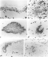
Photomicrographs of senile plaques from different brain regions, stained with a variety of methods from the author’s laboratory, including the Civin Laboratory modification of the Bielschowsky silver stain (A, B), thioflavin S (C), immunoperoxidase for Aβ with the 6E10 monoclonal antibody (D), Gallyas silver stain (E), and the Campbell-Switzer silver stain (F–L). Neuritic plaques are seen in (A) and (E), diffuse plaques in (B), and both types in (C) and (D). The Campbell-Switzer stain also shows plaques in the frontal cortex (F), putamen (G), cerebellar cortex (H), hippocampus (I), presubiculum and parasubiculum (J), entorhinal area (K) and calcarine cortex (L).
FIGURE 6.
FIGURE 6.
Frontal cortex sections demonstrating concordance between H3 PiB autoradiography (D) and 6E10 monoclonal antibody IHC for Aβ (F), from Lockhart et al (2007). Arrows demonstrate specific plaques marked by both PiB autoradiography and Aβ immunostaining. NDB = section treated with the nonradioactive competitive ligand BTA-1. Reprinted from Ref. (294), copyright Oxford University Press.
FIGURE 7.
FIGURE 7.
Paired F18 Florbetapir PET amyloid scans and Aβ immunohistochemistry from the same subjects after death. Reprinted from Ref. (299).
FIGURE 8.
FIGURE 8.
Negative (A) compared to highly positive (B) striatal F18 flutemetamol PET signal, in the coronal plane, with MR images for orientation. The striatal signal is centered over the gray matter of the head of the caudate nucleus and putamen (arrows). Reprinted from Ref. (313), with permission from IOS Press and the Journal of Alzheimer’s Disease.

References

    1. Khachaturian ZS. A chapter in the development of Alzheimer’s disease research: A case study of public policies on the development and funding of research programs. Alzheimers Dement 2007;3:243–258 - PubMed
    1. Selkoe DJ. Biochemistry and molecular biology of amyloid beta–protein and the mechanism of Alzheimer’s disease. Handb Clin Neurol 2008;89:245–60 - PubMed
    1. Nelson PT, Alafuzoff I, Bigio EH, et al. Correlation of Alzheimer disease neuropathologic changes with cognitive status: A review of the literature. J Neuropathol Exp Neurol 2012;71:362–81 - PMC - PubMed
    1. Terry RD, Katzman R.. Senile dementia of the Alzheimer type. Ann Neurol 1983;14:497–506 - PubMed
    1. Beach TG. Alzheimer disease and Down syndrome: Scientific symbiosis - a historical commentary. In: Berg JM, Karlinsky H, Holland J, eds. Alzheimer Disease, Down Syndrome and Their Relationship. Oxford: Oxford University Press; 1993:37–52

Publication types

MeSH terms

Substances