

Resting state functional brain networks associated with emotion processing in frontotemporal lobar degeneration
- PMID: 35595978
- PMCID: PMC9734056
- DOI: 10.1038/s41380-022-01612-9
Resting state functional brain networks associated with emotion processing in frontotemporal lobar degeneration
Abstract
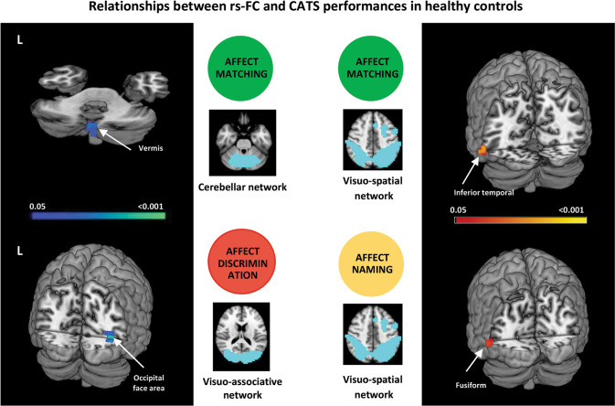
This study investigated the relationship between emotion processing and resting-state functional connectivity (rs-FC) of the brain networks in frontotemporal lobar degeneration (FTLD). Eighty FTLD patients (including cases with behavioral variant of frontotemporal dementia, primary progressive aphasia, progressive supranuclear palsy syndrome, motor neuron disease) and 65 healthy controls underwent rs-functional MRI. Emotion processing was tested using the Comprehensive Affect Testing System (CATS). In patients and controls, correlations were investigated between each emotion construct and rs-FC changes within critical networks. Mean rs-FC of the clusters significantly associated with CATS scoring were compared among FTLD groups. FTLD patients had pathological CATS scores compared with controls. In controls, increased rs-FC of the cerebellar and visuo-associative networks correlated with better scores in emotion-matching and discrimination tasks, respectively; while decreased rs-FC of the visuo-spatial network was related with better performance in the affect-matching and naming. In FTLD, the associations between rs-FC and CATS scores involved more brain regions, such as orbitofrontal and middle frontal gyri within anterior networks (i.e., salience and default-mode), parietal and somatosensory regions within visuo-spatial and sensorimotor networks, caudate and thalamus within basal-ganglia network. Rs-FC changes associated with CATS were similar among all FTLD groups. In FTLD compared to controls, the pattern of rs-FC associated with emotional processing involves a larger number of brain regions, likely due to functional specificity loss and compensatory attempts. These associations were similar across all FTLD groups, suggesting a common physiopathological mechanism of emotion processing breakdown, regardless the clinical presentation and pattern of atrophy.
© 2022. The Author(s).
Conflict of interest statement
EC receives or has received research supports form the Italian Ministry of Health; DC, VC, SB, MAM, NR, GM, FC, PC, SP, CV, DP, GM, LT, IA, and BP have nothing to disclose; VS received compensation for consulting services and/or speaking activities from AveXis, Cytokinetics and Italfarmaco; and receives or has received research supports form the Italian Ministry of Health, AriSLA, and E-Rare Joint Transnational Call; Prof. Filippi is Editor-in-Chief of the
Figures


Similar articles
-
Affective mentalizing and brain activity at rest in the behavioral variant of frontotemporal dementia.Neuroimage Clin. 2015 Aug 28;9:484-97. doi: 10.1016/j.nicl.2015.08.012. eCollection 2015. Neuroimage Clin. 2015. PMID: 26594631 Free PMC article.
-
MRI signatures of the frontotemporal lobar degeneration continuum.Hum Brain Mapp. 2015 Jul;36(7):2602-14. doi: 10.1002/hbm.22794. Epub 2015 Mar 28. Hum Brain Mapp. 2015. PMID: 25821176 Free PMC article.
-
Measuring disease progression in frontotemporal lobar degeneration: a clinical and MRI study.Neurology. 2010 Feb 23;74(8):666-73. doi: 10.1212/WNL.0b013e3181d1a879. Neurology. 2010. PMID: 20177120 Free PMC article.
-
Charting Frontotemporal Dementia: From Genes to Networks.J Neuroimaging. 2016 Jan-Feb;26(1):16-27. doi: 10.1111/jon.12316. Epub 2015 Nov 29. J Neuroimaging. 2016. PMID: 26617288 Review.
-
Atypical parkinsonian syndromes: a general neurologist's perspective.Eur J Neurol. 2018 Jan;25(1):41-58. doi: 10.1111/ene.13412. Epub 2017 Sep 28. Eur J Neurol. 2018. PMID: 28803444 Free PMC article. Review.
Cited by
-
A common marker of affect recognition dysfunction in the FTD spectrum of disorders.Eur J Neurol. 2025 Jan;32(1):e16578. doi: 10.1111/ene.16578. Eur J Neurol. 2025. PMID: 39632486 Free PMC article.
-
The predictive value of social cognition assessment for 1-year follow-up functional outcomes in behavioral variant frontotemporal dementia.J Neurol. 2025 Jul 21;272(8):526. doi: 10.1007/s00415-025-13254-2. J Neurol. 2025. PMID: 40691736
-
Changes in EEG Microstate Dynamics and Cognition Post-Chemotherapy in People With Breast Cancer.Brain Behav. 2025 Mar;15(3):e70335. doi: 10.1002/brb3.70335. Brain Behav. 2025. PMID: 40038798 Free PMC article. Clinical Trial.
-
Functional connectivity in behavioral variant frontotemporal dementia.Brain Behav. 2022 Dec;12(12):e2790. doi: 10.1002/brb3.2790. Epub 2022 Oct 28. Brain Behav. 2022. PMID: 36306386 Free PMC article. Review.
-
Dual-task gait training improves cognition and resting-state functional connectivity in Parkinson's disease with postural instability and gait disorders.J Neurol. 2024 Apr;271(4):2031-2041. doi: 10.1007/s00415-023-12151-w. Epub 2024 Jan 8. J Neurol. 2024. PMID: 38189921 Clinical Trial.
References
-
- Ekman P, Friesen WV. Constants across cultures in the face and emotion. J Pers Soc Psychol. 1971;17:124–9. - PubMed
-
- Ekman P. Pictures of facial affect. Consulting Psychologists Press; Palo Alto, CA, 1976.
-
- Oliver LD, Mitchell DG, Dziobek I, MacKinley J, Coleman K, Rankin KP, et al. Parsing cognitive and emotional empathy deficits for negative and positive stimuli in frontotemporal dementia. Neuropsychologia. 2015;67:14–26. - PubMed
-
- Piguet O, Leyton CE, Gleeson LD, Hoon C, Hodges JR. Memory and emotion processing performance contributes to the diagnosis of non-semantic primary progressive aphasia syndromes. J Alzheimer’s Dis: JAD. 2015;44:541–7. - PubMed
