. 2022 Nov;29(11):1628-1635.
doi: 10.1038/s41417-022-00480-3. Epub 2022 May 20.

Ixovex-1, a novel oncolytic E1B-mutated adenovirus

Affiliations

Ixovex-1, a novel oncolytic E1B-mutated adenovirus

Mohiemen Anwar et al. Cancer Gene Ther. 2022 Nov.

Abstract

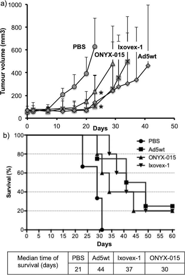
There is a great demand for improved oncolytic viruses that selectively replicate within cancer cells while sparing normal cells. Here, we describe a novel oncolytic adenovirus, Ixovex-1, that obtains a cancer-selective replication phenotype by modulating the level of expression of the different, alternatively spliced E1B mRNA isoforms. Ixovex-1 is a recombinant adenovirus that carries a single point mutation in the E1B-93R 3' splice acceptor site that results in overexpression of the E1B-156R splice isoform. In this paper, we studied the characteristics of this novel oncolytic adenovirus by validating its in vitro behaviour in a panel of normal cells and cancer cells. We additionally studied its anti-tumour efficacy in vivo. Ixovex-1 significantly inhibited tumour growth and prolonged survival of mice in an immune-deficient lung carcinoma tumour implantation model. In complementation experiments, overexpression of E1B-156R was shown to increase the oncolytic index of both Ad5wt and ONYX-015. In contrast to prior viruses of similar type, Ixovex-1 includes a functional E3B region for better in vivo efficacy. Throughout this study, the Ixovex-1 virus has been proven to be superior in competency compared to a virus with multiple deletions.


Conflict of interest statement

AC, GA, M-LA, MR and ME have no conflict of interests. DÖ and MA have both stocks in Ixogen Ltd., UK. GhA is the founder and CEO of Ixogen Ltd. Ixogen holds worldwide patents to Ixovex-1. GhA is also the founder and CEO of PsiVac Ltd., UK.

Figures

Fig. 1
Fig. 1. The adenovirus genome and E1B cassette.
a The full-length genome with the E1 and E3 gene cassettes is indicated. b E1B splice map showing alternative E1B gene products. The full-length E1B transcript carries the E1B-176R, E1B-496R and pIX ORF. Through alternative splicing within the E1B-496R ORF, another three additional mRNAs are produced encoding for proteins, E1B-93R, E1B-156R and E1B-84R. The spliced E1B products have the 79 N-terminal aa in common with E1B-496R but differ in the C-terminal, except for E1B-156R, which splices in frame with E1B-496R.
Fig. 2
Fig. 2. An amino acid change in the E1B-496R protein results in a loss of protein accumulation.
A549 cells were infected with the respective virus at 2.5 pfu/cell and total cell lysate was collected at 48 hpi. Shown is a western blot stained with polyclonal α-capsid (late) protein antibody (top panel), a monoclonal α-E1B-496R antibody (2A6, middle panel) and a monoclonal α-actin antibody as a loading control (lowest panel). The Control lane is lysate from non-infected cells.
Fig. 3
Fig. 3. The point mutation in the E1B-496R ORF in Ixovex-1 blocks splicing to the E1B-93R splice acceptor (SA1).
A549 cells were infected with the respective virus at 2.5 pfu/cell and a total RNA was collected at 48 hpi. cDNA was made using an oligo-dT oligonucleotide. PCR was performed using a common sense oligonucleotide upstream of the splice donor 1 and a specific antisense oligonucleotide downstream of the respective splice acceptor. The PCR reactions were run on a 2% agarose gel. b In parallel, total protein was harvested at 48 hpi and western blot was performed using the 2A6 antibody that binds the common N-terminus shared by E1B-496R and its smaller spliced versions, an anti-E1A and a polyclonal anti-capsid (late) protein antibody.
Fig. 4
Fig. 4. Ixovex-1 fails to induce p53 degradation, but does not induce p53 accumulation.
A549 cells were infected with the respective virus at 2.5 pfu/cell and total cell lysate was collected 48 hpi. Shown is a western blot using a monoclonal α-p53 antibody (top panel) and the monoclonal α-actin antibody as a loading control (lower panel). Mock lane is lysate from non-infected cells.
Fig. 5
Fig. 5. Replication-efficiencies in H460 and H1299 cancer cells.
Each cell line was infected with 2.5 pfu/cell of the respective virus. Cells and media were harvested at 24, 48 and 72 hpi and analysed by a limited dilution assay. CPE was scored after 10 days and TCID50 (pfu/cell) results were calculated.
Fig. 6
Fig. 6. Replication-competencies in NHBE.
Cells were infected with 1 pfu/cell of the respective virus. Cells and media were harvested at 24, 48 and 72 hpi and analysed by a limited dilution assay. CPE was scored after 10 days and TCID50 (pfu/cell) results were calculated. The result for Ixovex-1 was adjusted upwards because the level of replication was slightly below the detection limit.
Fig. 7
Fig. 7. Ixovex-1 significantly inhibits tumour growth in nude mice and prolongs survival.
Nude NMRI mice were injected subcutaneously with H1299 cells in PBS mixed 1:1 with matrigel. After 2 weeks, mice with tumour growth (size ~70–100 mm3) were randomised into four groups with seven mice in each. Tumours were injected on days -2, -1, and 0 with (108 viral particles) Ad5wt, ONYX-015, Ixovex-1 or PBS only. a All tumours were measured using a digital caliper two times per week. Tumour size was calculated as: length × width × depth × 0.66. Mice were sacrificed when tumour size exceeded 1000 mm3. Data plotted are the volume means ± SEM. One-way ANOVA analysis of variance was performed comparing the growth curves. Prism 6 software (GraphPad Software Inc.) was used throughout the analysis. b The proportions of live mice are plotted against time. The median time of survival was determined by Kaplan–Meier survival analysis (*p < 0.05, log-rank test for statistical significance).

Similar articles

Cited by

References

    1. Cheng PH, Wechman SL, McMasters KM, Zhou HS. Oncolytic replication of E1b-deleted adenoviruses. Viruses. 2015;7:5767–79. doi: 10.3390/v7112905. - DOI - PMC - PubMed
    1. Turnbull S, West EJ, Scott KJ, Appleton E, Melcher A, Ralph C. Evidence for oncolytic virotherapy: where have we got to and where are we going? Viruses. 2015;7:6291–312. doi: 10.3390/v7122938. - DOI - PMC - PubMed
    1. Uusi-Kerttula H, Hulin-Curtis S, Davies J, Parker AL. Oncolytic adenovirus: strategies and insights for vector design and immuno-oncolytic applications. Viruses. 2015;7:6009–42. doi: 10.3390/v7112923. - DOI - PMC - PubMed
    1. Wang X, Simpson ER, Brown KA. p53: protection against tumor growth beyond effects on cell cycle and apoptosis. Cancer Res. 2015;75:5001–7. doi: 10.1158/0008-5472.CAN-15-0563. - DOI - PubMed
    1. Moore PS, Chang Y. Why do viruses cause cancer? Highlights of the first century of human tumour virology. Nat Rev Cancer. 2010;10:878–89. doi: 10.1038/nrc2961. - DOI - PMC - PubMed

Publication types

Substances