. 2022 May 20;22(1):567.
doi: 10.1186/s12885-022-09666-2.

Mir-675-5p supports hypoxia-induced drug resistance in colorectal cancer cells

Affiliations

Mir-675-5p supports hypoxia-induced drug resistance in colorectal cancer cells

Chiara Zichittella et al. BMC Cancer. .

Abstract

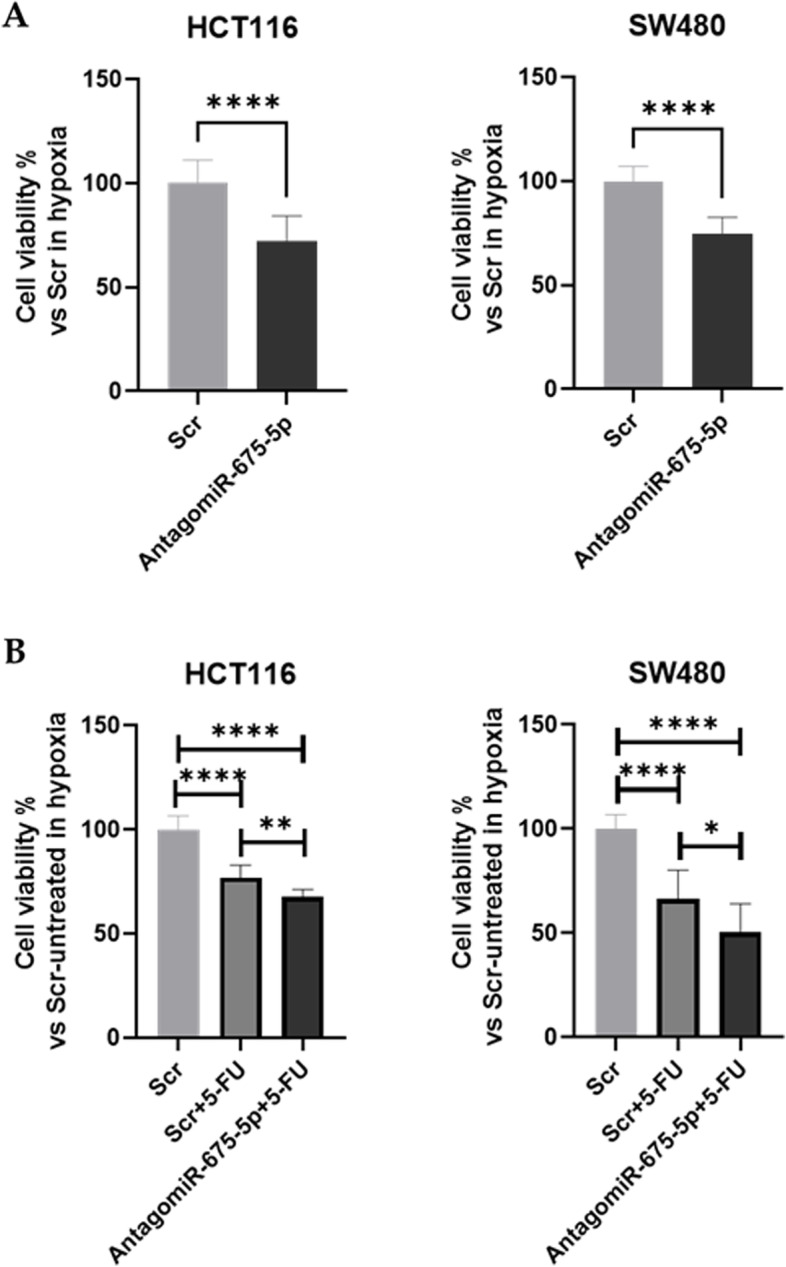
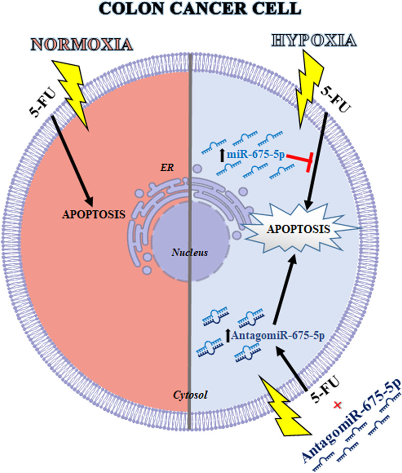
Background: The uncontrolled proliferation of cancer cells determines hypoxic conditions within the neoplastic mass with consequent activation of specific molecular pathways that allow cells to survive despite oxygen deprivation. The same molecular pathways are often the cause of chemoresistance. This study aims to investigate the role of the hypoxia-induced miR-675-5p in 5-Fluorouracil (5-FU) resistance on colorectal cancer (CRC) cells.

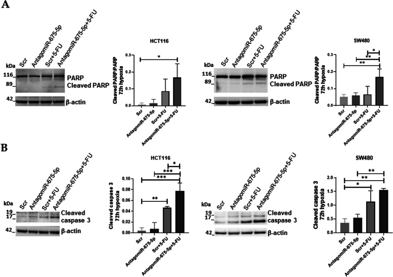
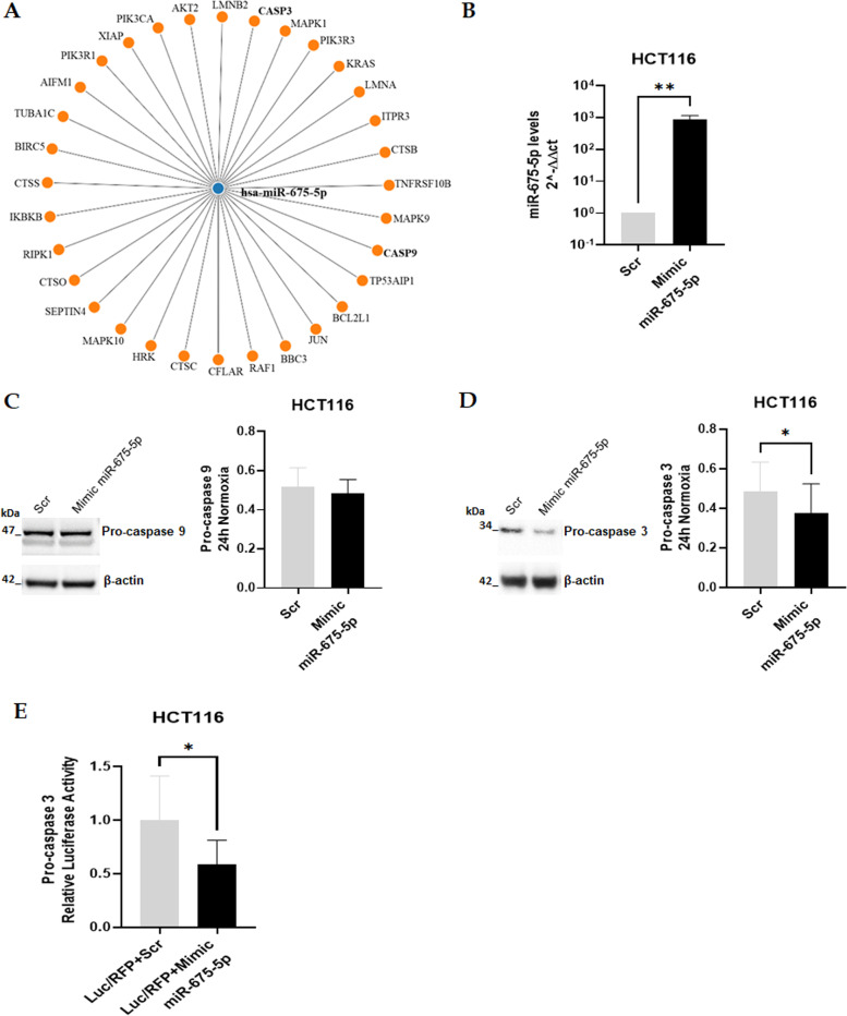
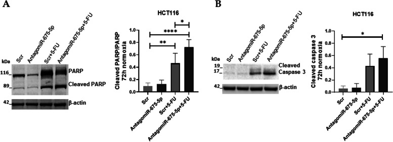
Methods: CRC cell lines were treated with 5-Fu and incubated in normoxic or hypoxic conditions; cell viability has been evaluated by MTT assay. MiR-675-5p levels were analysed by RT-PCR and loss and gain expression of the miRNA has been obtained by the transfection of miRNA antagomir or miRNA mimic. Total protein expression of different apoptotic markers was analysed through western blot assay. MirWalk 2.0 database search engine was used to investigate the putative targets of the miR-675-5p involved in the apoptotic process. Finally, the luciferase assay was done to confirm Caspase-3 as a direct target of the miR-675-5p.

Results: Our data demonstrated that hypoxia-induced miR-675-5p counteracts the apoptotic signal induced by 5-FU, thus taking part in the drug resistance response. We showed that the apoptotic markers, cleaved PARP and cleaved caspase-3, increased combining miR-675-5p inhibition with 5-FU treatment. Moreover, we identified pro-caspase-3 among the targets of the miR-675-5p.

Conclusion: Our data demonstrate that the inhibition of hypoxia-induced miR-675-5p combined with 5-FU treatment can enhances drug efficacy in both prolonged hypoxia and normoxia, indicating a possible strategy to partially overcome chemoresistance.

Keywords: 5-fluorouracil (5-FU); Apoptosis; Colorectal cancer (CRC); Drug resistance; Hypoxia; MicroRNA.


Conflict of interest statement

The authors declare that they have no competing interests

Figures

Fig. 1
Fig. 1
Carbonic Anhydrase 9 expression confirms the hypoxic condition. A-B: Representative images and densitometric analysis of Western blots for Carbonic Anhydrase (CA9) obtained from protein lysates of HCT116 and SW480 in normoxic conditions or subjected to hypoxic conditions. The graphs ordinate shows the OD (Optical Density) of the indicated proteins normalized for the housekeeping’s OD (β-actin). Data are expressed as the mean ± SD of three independent experiments and statistical significance was analyzed using a Student’s t-test (* = p < 0.05; ** = p < 0.01). Corresponding uncropped full-length blots are included in Supplementary Materials
Fig. 2
Fig. 2
Colon cancer cell lines behaviour under chronic hypoxic stimulation (72 h). A-B: Cell viability assay (MTT Assay) in HCT116 and SW480 treated for 72 h with two different concentrations of 5-FU (5 μM and 10 μM) in normoxic (N) conditions or subjected to hypoxic (H) conditions. Data are expressed as the percentage of cell viability versus untreated cells both in normoxia and chronic hypoxia (Ctr). C: Analysis of the expression level (qRT-PCR) of miR-675-5p in HCT116 and SW480 under normoxic conditions and after 72 hours of hypoxic stimulation. The miR-675-5p levels were normalized for RNU6 (U6 Small Nuclear 1), and the ΔΔCt was calculated with respect to the expression levels under normoxic conditions. All data are the mean ± SD of three biological replicates. Statistical analyses: Ordinary one-way ANOVA with Bonferroni’s multiple comparison test were used in Figs. A and B, Student’s t-test was used for Fig. C (* = p < 0.05; ** = p < 0.01; **** = p < 0.0001)
Fig. 3
Fig. 3
Effects of AntagomiR-675-5p treatment in cell viability in chronic hypoxic conditions. A: Cell viability assay (MTT Assay) in HCT116 and SW480 transfected with AntagomiR-675-5p or Scrambled Negative Control (Scr) and grown in the hypoxic chamber for 72 h. Data are expressed as cell viability percentage compared to cells transfected with Scr. B: Cell viability assay (MTT Assay) in HCT116 and SW480 transfected with AntagomiR-675-5p or Scramble Negative Control (Scr) and treated or not for 72 h of hypoxia with 5-FU (10 μM). Data are expressed as the mean ± SD of three biological replicates. Statistical analyses: Student’s t-test was used for Fig. A, Ordinary one-way ANOVA with Bonferroni’s multiple comparison test were used in Fig. B (* = p < 0.05; ** = p < 0.01; **** = p < 0.0001)
Fig. 4
Fig. 4
Effects of AntagomiR-675-5p treatment on apoptosis markers. A-B: Representative images and densitometric analysis of Western blots for cleaved PARP/PARP and Cleaved caspase-3 obtained from protein lysates of HCT116 and SW480 in chronic hypoxia, transfected with AntagomiR-675-5p or Scrambled Negative Control (Scr) and treated or not with 5-FU (10 μM). The graphs ordinate shows the OD (Optical Density) of the indicated proteins normalized for the housekeeping’s OD (β-actin). Data are expressed as the mean ± SD of three independent experiments and statistical significance was analyzed by using Ordinary one-way ANOVA with Bonferroni’s multiple comparison test (* = p < 0.05; ** = p < 0.01; *** = p < 0.001; **** = p < 0.0001). Corresponding uncropped full-length blots are included in a Supplementary Material
Fig. 5
Fig. 5
Identification of miR-675-5p targets involved in apoptosis. A: The network diagram obtained using the mirWalk database [29] illustrates the presumed 3’UTR targets of miR-675-5p involved in apoptosis (KEGG Pathway hsa04210#Apoptosis). B: Expression level analysis (qRT-PCR) of miR-675-5p in HCT116 after overexpression of miR-675-5p under normoxic conditions. The miR-675-5p levels were normalized for RNU6 (U6 Small Nuclear 1), and the ΔΔCt was calculated with respect to the Scrambled Negative Control (Scr). C-D: Representative images and densitometric analysis of Western blots respectively for pro-caspase 9 and pro-caspase 3 on proteins lysates from HCT116 transfected with miR-675-5p mimic or Scrambled Negative Control (Scr) for 24 h in normoxia. The graphs ordinate shows the OD (Optical Density) of the indicated proteins normalized for the housekeeping’s OD (β-actin). Corresponding uncropped full-length blots are included in a Supplementary Materials. E: The Firefly Luciferase assay validates pro-caspase 3 as the target of miR-675-5p. Luminescence was normalized for RFP values end presented in the graph as relative Luciferase activity in cells treated with mimic-miR-675-5p (Luc/RFP + Mimic miR-675-5p) with respect to cells treated with the Negative Control (Luc/RFP + Scr). All Data are expressed as mean ± SD of three independent experiments and statistical significance was analyzed using Student’s t-test (* = p < 0.05; ** = p < 0.01)
Fig. 6
Fig. 6
Effects of AntagomiR-675-5p on HCT116 cell line in normoxic conditions. A-B: Representative images and densitometric analyses of Western blots for cleaved PARP/PARP and Cleaved caspase-3 in HCT116 in normoxic conditions, transfected with AntagomiR-675-5p or Scrambled Negative Control (Scr) and treated or not with 5-FU (10 μM). The graphs ordinate shows the OD (Optical Density) of the indicated proteins normalized for the housekeeping’s OD (β-actin). Data are expressed as the mean ± SD of three independent experiments and statistical significance was analysed by using Ordinary one-way ANOVA with Bonferroni’s multiple comparison test (* = p < 0.05; ** = p < 0.01; **** = p < 0.0001). Corresponding uncropped full-length blots are included in a Supplementary Material
Fig. 7
Fig. 7
Schematic representation of the proposed model. On the left in red is represented the CRC cell treated with the chemotherapeutic drug 5-Fluorouracil (5-FU) which in normoxic conditions activates the apoptotic process. In blue at the top right is represented the CRC cell treated with 5-FU in conditions of prolonged hypoxia, in which the overexpression of miR-675-5p inhibits the activation of the apoptotic process by targeting the pro-caspase 3. Finally, below on the right in blue is represented the CRC cell treated with 5-FU in conditions of prolonged hypoxia, in which the presence of AntagomiR-675-5p activates the apoptotic process, increasing the protein levels of the cleaved caspase-3 and the cleaved PARP

Similar articles

Cited by

References

    1. Rawla P, Sunkara T, Barsouk A. Epidemiology of colorectal cancer: incidence, mortality, survival, and risk factors. Prz Gastroenterol. 2019;14(2):89–103. - PMC - PubMed
    1. Dekker E, Tanis PJ, Vleugels JLA, Kasi PM, Wallace MB. Colorectal cancer. Lancet. 2019;394(10207):1467–1480. doi: 10.1016/S0140-6736(19)32319-0. - DOI - PubMed
    1. Thanikachalam K, Khan G. Colorectal Cancer and nutrition. Nutrients. 2019;11:1. doi: 10.3390/nu11010164. - DOI - PMC - PubMed
    1. Buccafusca G, Proserpio I, Tralongo AC, Rametta Giuliano S, Tralongo P. Early colorectal cancer: diagnosis, treatment and survivorship care. Crit Rev Oncol Hematol. 2019;136:20–30. doi: 10.1016/j.critrevonc.2019.01.023. - DOI - PubMed
    1. Francipane MG, Bulanin D, Lagasse E. Establishment and characterization of 5-fluorouracil-resistant human colorectal Cancer stem-like cells: tumor dynamics under selection pressure. Int J Mol Sci. 2019;20(8). - PMC - PubMed