. 2022 May 20;9(1):22.
doi: 10.1186/s40779-022-00381-4.

Celastrol mitigates inflammation in sepsis by inhibiting the PKM2-dependent Warburg effect

Affiliations

Celastrol mitigates inflammation in sepsis by inhibiting the PKM2-dependent Warburg effect

Piao Luo et al. Mil Med Res. .

Abstract

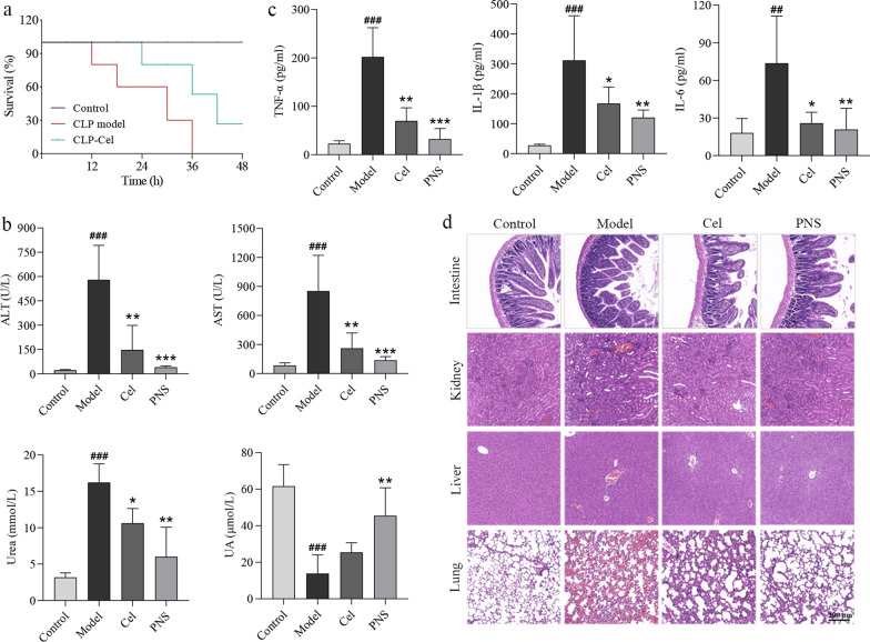
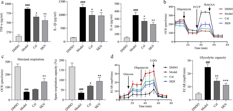
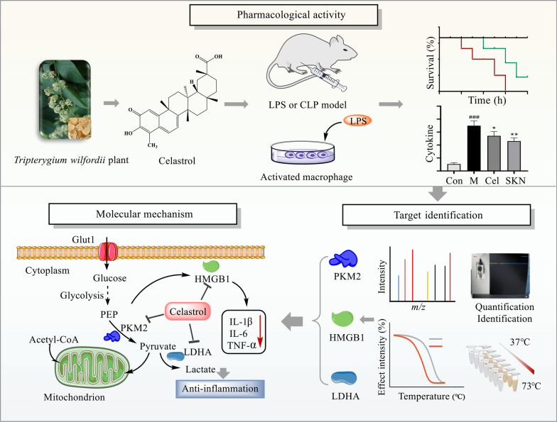
Background: Sepsis involves life-threatening organ dysfunction and is caused by a dysregulated host response to infection. No specific therapies against sepsis have been reported. Celastrol (Cel) is a natural anti-inflammatory compound that shows potential against systemic inflammatory diseases. This study aimed to investigate the pharmacological activity and molecular mechanism of Cel in models of endotoxemia and sepsis.

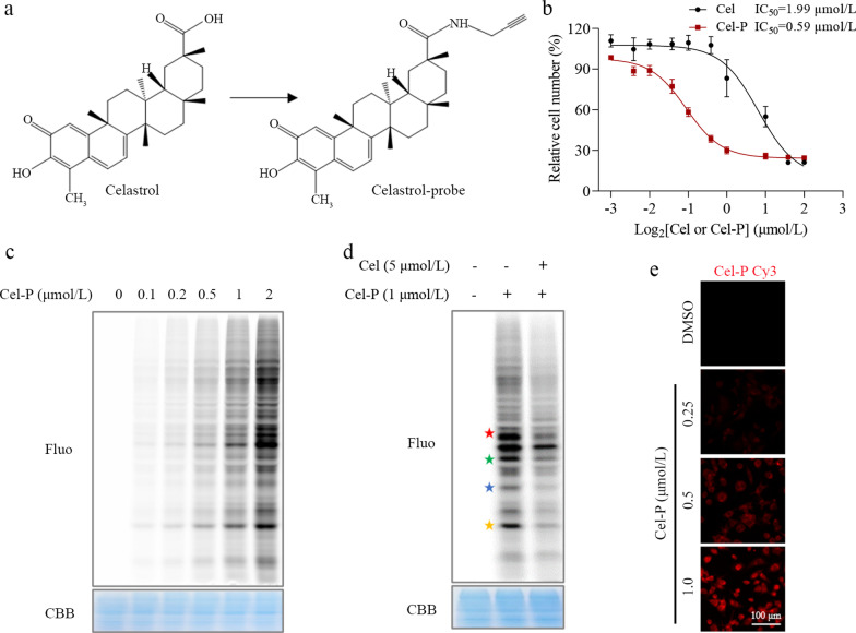
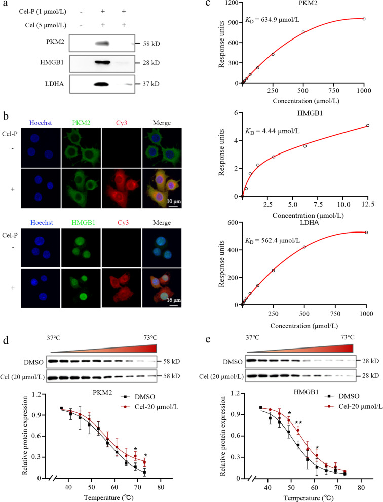
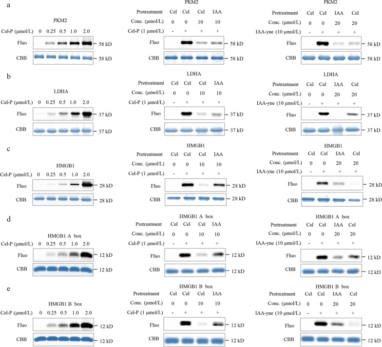
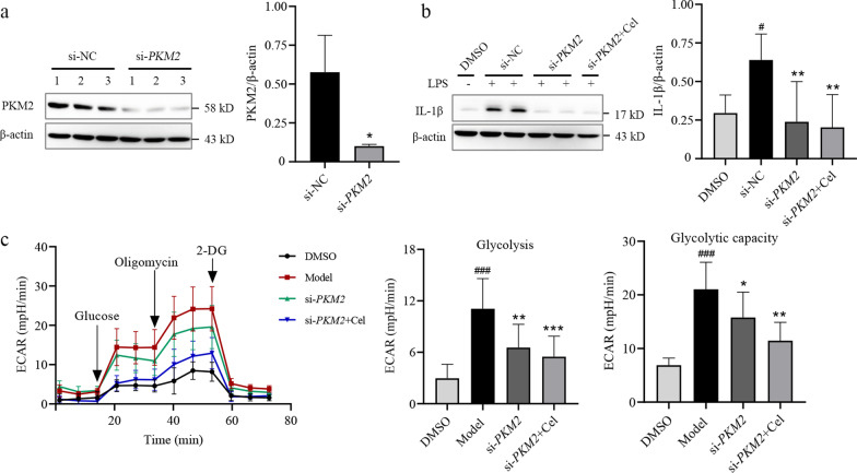
Methods: We evaluated the anti-inflammatory efficacy of Cel against endotoxemia and sepsis in mice and macrophage cultures treated with lipopolysaccharide (LPS). We screened for potential protein targets of Cel using activity-based protein profiling (ABPP). Potential targets were validated using biophysical methods such as cellular thermal shift assays (CETSA) and surface plasmon resonance (SPR). Residues involved in Cel binding to target proteins were identified through point mutagenesis, and the functional effects of such binding were explored through gene knockdown.

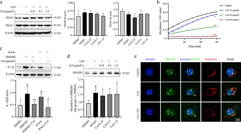
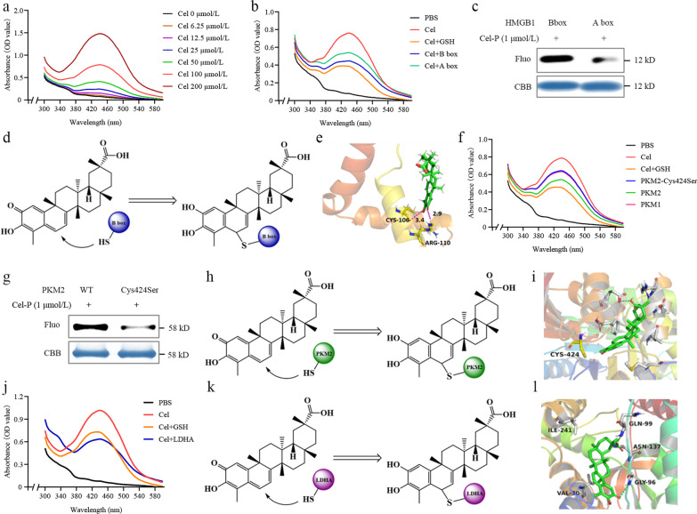
Results: Cel protected mice from lethal endotoxemia and improved their survival with sepsis, and it significantly decreased the levels of pro-inflammatory cytokines in mice and macrophages treated with LPS (P < 0.05). Cel bound to Cys424 of pyruvate kinase M2 (PKM2), inhibiting the enzyme and thereby suppressing aerobic glycolysis (Warburg effect). Cel also bound to Cys106 in high mobility group box 1 (HMGB1) protein, reducing the secretion of inflammatory cytokine interleukin (IL)-1β. Cel bound to the Cys residues in lactate dehydrogenase A (LDHA).

Conclusion: Cel inhibits inflammation and the Warburg effect in sepsis via targeting PKM2 and HMGB1 protein.

Keywords: Aerobic glycolysis; Celastrol; High mobility group box 1; Pyruvate kinase M2; Sepsis.


Conflict of interest statement

The authors declare no conflicts of interest.

Figures

Fig. 1
Fig. 1
Celastrol (Cel) protects mice from experimental sepsis and endotoxic shock. a Survival rates of cecal ligation puncture (CLP) mice with or without Cel (1 mg/kg, i.p.). b Serum indicators of liver and kidney function in endotoxemic mice. c Release of the inflammatory cytokines TNF-α, IL-1β and IL-6 in endotoxemic mice. d Hematoxylin–eosin staining of intestine, kidney, liver and lung sections from endotoxemic mice. Scale bar = 200 μm. All data are expressed as mean ± SEM (n = 6). ###P < 0.001 vs. Control; *P < 0.05, **P < 0.01, ***P < 0.001 vs. Model. ALT alanine aminotransferase, PNS prednisone, AST aspartate aminotransferase, UA uric acid, TNF-α tumor necrosis factor-α, IL-1β interleukin-1β, IL-6 interleukin-6
Fig. 2
Fig. 2
Celastrol (Cel) inhibits inflammatory response by suppressing the Warburg effect in LPS-activated macrophages. The grouping as follows: DMSO, Model (LPS 100 ng/ml), Cel (LPS 100 ng/ml + Cel 1 μmol/L), SKN (LPS 100 ng/ml + shikonin 1 μmol/L). a Release of the pro-inflammatory cytokines TNF-α, IL-1β and IL-6. b Oxygen consumption rate (OCR). c Maximal respiration and spare respiration capacity of OCR. d Extracellular acidification rate (ECAR). All data are expressed as mean ± SEM (n = 3). ###P < 0.001 vs. DMSO; *P < 0.05, **P < 0.01, ***P < 0.001 vs. Model. LPS lipopolysaccharide, SKN shikonin, TNF-α tumor necrosis factor-α, IL-1β interleukin-1β, IL-6 interleukin-6, FCCP p-trifluoromethoxy carbonyl cyanide phenylhydrazone, Rote/AA rotenone/antimycin A, 2-DG 2-deoxy-glucose
Fig. 3
Fig. 3
Identification of potential target proteins of celastrol (Cel) through ABPP combined with LC–MS/MS. a Chemical structure of Cel and celastrol-probe (Cel-P). b Dose-dependent inhibition of RAW264.7 cell proliferation by Cel and Cel-P. c Dose-dependent labeling of proteins by Cel-P in LPS-activated macrophages. d Competition between Cel and Cel-P for protein binding in situ (red star: 58 kD, green star: 43 kD, blue star: 37 kD, yellow star: 25 kD). e Imaging of intracellular Cel-P. Scale bar = 100 μm. ABPP activity-based protein profiling, LC–MS/MS liquid chromatography – tandem mass spectrometry, Fluo fluorescence, CBB coomassie brilliant blue
Fig. 4
Fig. 4
Celastrol (Cel) directly targets PKM2 and HMGB1. a Western blotting of pull-downs to verify Cel binding to PKM2, HMGB1 and LDHA in situ. b Immunofluorescence staining of PKM2 and HMGB1 (green) and Cel-P click-conjugated to a TAMRA dye (red). Scale bar = 10 μm or 12 μm. c Surface plasmon resonance studies of the binding affinity of Cel with recombinant human PKM2, HMGB1 and LDHA. d Cellular thermal shift assays to verify Cel binding to PKM2. e Cellular thermal shift assays to verify Cel binding to HMGB1 (n = 3). *P < 0.05, **P < 0.01 vs. 37 °C. Cel-P celastrol-probe, PKM2 pyruvate kinase M2, HMGB1 high mobility group box 1, LDHA lactate dehydrogenase A
Fig. 5
Fig. 5
Celastrol (Cel) suppresses the enzymatic activity of pyruvate kinase and the proinflammatory activity of HMGB1. a Western blotting against PKM1 and PKM2 in macrophages treated or not with Cel at 0.25, 0.5 or 1.0 μmol/L. The corresponding densitometry is shown (n = 3). b Catalytic activity of pyruvate kinase with or without Cel treatment at 10, 20 and 40 μmol/L, n = 3, *P < 0.05, **P < 0.01, ***P < 0.001 vs. DMSO. c The contribution of HMGB1 to IL-1β expression, as determined by Western blotting. Assays were set up with HMGB1 at 0.8 μg/ml; B box, 0.8 μg/ml; and Cel, 0.2 μmol/L (n = 3). #P < 0.05 vs. DMSO; *P < 0.05, **P < 0.01 vs. HMGB1 or B box. d Secretion of HMGB1 from macrophages treated or not with Cel at 0.25, 0.5 or 1.0 μmol/L, as determined using Western blotting (n = 3). #P < 0.05 vs. DMSO. e Immunofluorescence staining with antibody against HMGB1 (green) or a dye targeting the cell membrane (red). Scale bar = 6 μm. LPS lipopolysaccharide, PKM2 pyruvate kinase M2, PKM1 pyruvate kinase M1, HMGB1 high mobility group box 1, IL-1β interleukin-1β
Fig. 6
Fig. 6
Celastrol (Cel) binds to Cys residues in PKM2, LDHA and HMGB1. a Recombinant human PKM2 labeled by celastrol-probe (Cel-P) or alkyne-tagged IAA in the presence or absence of the competitors Cel or IAA. b Recombinant human LDHA labeled by celastrol-probe (Cel-P) or alkyne-tagged IAA in the presence or absence of the competitors Cel or IAA. c Recombinant human HMGB1 labeled by celastrol-probe (Cel-P) or alkyne-tagged IAA in the presence or absence of the competitors Cel or IAA. d Recombinant human HMGB1 A box labeled by celastrol-probe (Cel-P) or alkyne-tagged IAA in the presence or absence of the competitors Cel or IAA. e Recombinant human HMGB1 B box labeled by celastrol-probe (Cel-P) or alkyne-tagged IAA in the presence or absence of the competitors Cel or IAA. PKM2 pyruvate kinase M2, LDHA lactate dehydrogenase A, HMGB1 high mobility group box 1, Conc. concentration, Fluo fluorescence, CBB coomassie brilliant blue, IAA iodoacetamide
Fig. 7
Fig. 7
Celastrol (Cel) reacts with Cys residues in PKM2 and HMGB1 to inhibit their activities. a Absorption spectra of Cel at 6.25–200 μmol/L. b Absorption spectra of 100 μmol/L Cel in the presence or absence of GSH, HMGB1 A box, or B box. c Fluorescence intensity of recombinant A box and B box incubated with celastrol-probe (Cel-P) and then click-reacted with a fluorescent dye. d Scheme depicting Cel binding to B box. e Molecular docking model of Cel binding to HMGB1. f Absorption spectra of 100 μmol/L Cel in the presence or absence of GSH, PKM1, wild-type PKM2, or PKM2-Cys424Ser. g Fluorescence intensity of recombinant wild-type PKM2 and PKM2-Cys424Ser incubated with Cel-P and then click-reacted with a fluorescent dye. h Scheme depicting Cel binding to PKM2. i Molecular docking model of Cel binding to PKM2. j Absorption spectra of 100 μmol/L Cel in the presence or absence of GSH and LDHA. k Scheme of Cel binding to LDHA. l Molecular ducking model of Cel binding to LDHA. PBS phosphate-buffered saline, GSH glutathione, LDHA lactate dehydrogenase A, PKM2 pyruvate kinase M2, HMGB1 high mobility group box 1, Fluo fluorescence, CBB coomassie brilliant blue
Fig. 8
Fig. 8
Celastrol (Cel) attenuates lipopolysaccharide (LPS)-induced inflammatory response and glycolysis. a Transfection of macrophages with short interfering (si)RNA against PKM2, and Western blotting with densitometry (n = 3). *P < 0.05 vs. si-NC. b IL-1β levels in cells transfected with si-PKM2 or si-NC and cultured in the presence or absence of Cel at 0.5 μmol/L (n = 3). #P < 0.05 vs. DMSO; **P < 0.01 vs. si-NC. c ECAR of cells transfected with si-PKM2 in the presence or absence of Cel at 0.5 μmol/L (n = 3). ###P < 0.001 vs. DMSO; *P < 0.05, **P < 0.01, ***P < 0.001 vs. Model. PKM2 pyruvate kinase M2, IL-1β interleukin-1β, ECAR extracellular acidification rate, 2-DG 2-deoxy-glucose, NC negative control
Fig. 9
Fig. 9
Summary of the key findings in this paper. Celastrol (Cel) protects mice from experimental sepsis and endotoxic shock by inhibiting secretion of pro-inflammatory cytokines. Cel directly targets PKM2, HMGB1, and LDHA, as shown using activity-based protein profiling and cellular thermal shift assays. Cel dampens the inflammatory response and glycolysis in LPS-activated macrophages by repressing the activities of PKM2 and HMGB1. LPS lipopolysaccharide, CLP cecal ligation puncture, PEP phosphor-enol-pyruvate, PKM2 pyruvate kinase M2, HMGB1 high mobility group box 1, LDHA lactate dehydrogenase A, IL-1β interleukin-1β, IL-6 interleukin-6, TNF-α tumor necrosis factor-α

Similar articles

Cited by

References

    1. Sygitowicz G, Sitkiewicz D. Molecular mechanisms of organ damage in sepsis: an overview. Braz J Infect Dis. 2020;24(6):552–560. doi: 10.1016/j.bjid.2020.09.004. - DOI - PMC - PubMed
    1. Xu H, Turnquist HR, Hoffman R, Billiar TR. Role of the IL-33-ST2 axis in sepsis. Mil Med Res. 2017;4:3. - PMC - PubMed
    1. Dreschers S, Ohl K, Lehrke M, Möllmann J, Denecke B, Costa I, et al. Impaired cellular energy metabolism in cord blood macrophages contributes to abortive response toward inflammatory threats. Nat Commun. 2019;10(1):1685. doi: 10.1038/s41467-019-09359-8. - DOI - PMC - PubMed
    1. Qiu D, Chu X, Hua L, Yang Y, Li K, Han Y, et al. Gpr174-deficient regulatory T cells decrease cytokine storm in septic mice. Cell Death Dis. 2019;10(3):233. doi: 10.1038/s41419-019-1462-z. - DOI - PMC - PubMed
    1. Wong TL, Ng KY, Tan KV, Chan LH, Zhou L, Che N, et al. CRAF methylation by PRMT6 regulates aerobic glycolysis-driven hepatocarcinogenesis via ERK-dependent PKM2 nuclear relocalization and activation. Hepatology. 2020;71(4):1279–1296. doi: 10.1002/hep.30923. - DOI - PubMed

Publication types