. 2022 May 20;21(1):31.
doi: 10.1186/s12938-022-01000-y.

Three-dimensional finite element analysis of the effect of alveolar cleft bone graft on the maxillofacial biomechanical stabilities of unilateral complete cleft lip and palate

Affiliations

Three-dimensional finite element analysis of the effect of alveolar cleft bone graft on the maxillofacial biomechanical stabilities of unilateral complete cleft lip and palate

Tao Tian et al. Biomed Eng Online. .

Abstract

Background: The objective is to clarify the effect of alveolar cleft bone graft on maxillofacial biomechanical stabilities, the key areas when bone grafting and in which should be supplemented with bone graft once bone resorption occurred in UCCLP (unilateral complete cleft lip and palate).

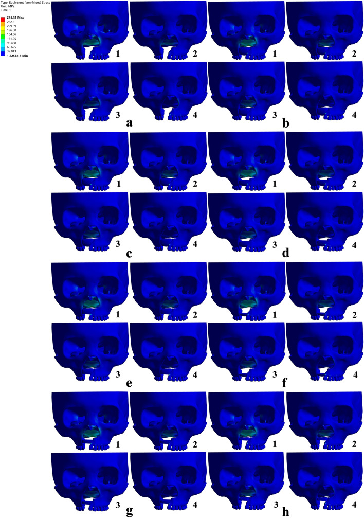
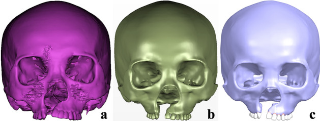
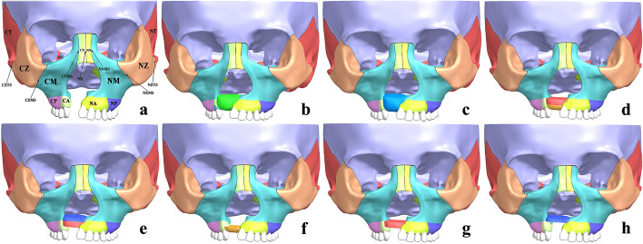
Methods: Maxillofacial CAD (computer aided design) models of non-bone graft and full maxilla cleft, full alveolar cleft bone graft, bone graft in other sites of the alveolar cleft were acquired by processing the UCCLP maxillofacial CT data in three-dimensional modeling software. The maxillofacial bone EQV (equivalent) stresses and bone suture EQV strains under occlusal states were obtained in the finite element analysis software.

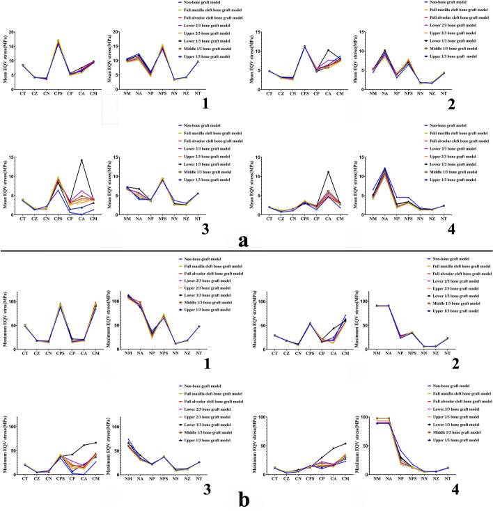
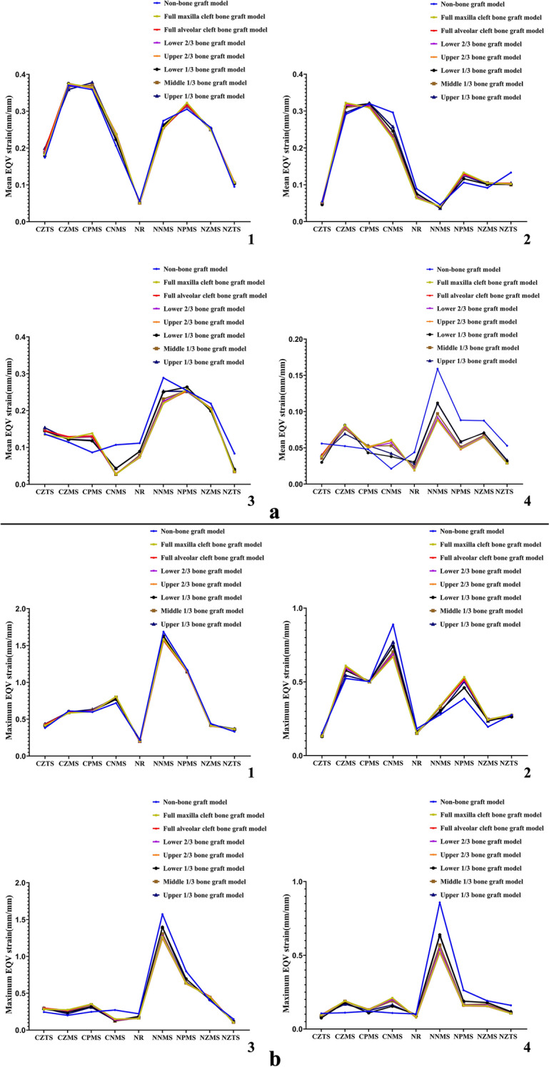
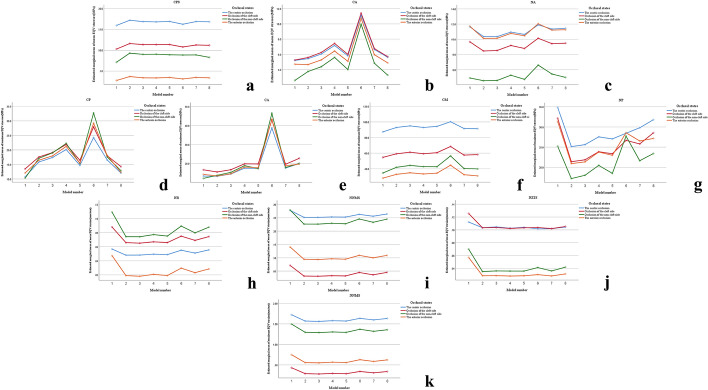
Results: Under corresponding occlusal states, the EQV stresses of maxilla, pterygoid process of sphenoid bone on the corresponding side and anterior alveolar arch on the non-cleft side were higher than other maxillofacial bones, the EQV strains of nasomaxillary, zygomaticomaxillary and pterygomaxillary suture on the corresponding side were higher than other maxillofacial bone sutures. The mean EQV strains of nasal raphe, the maximum EQV stresses of posterior alveolar arch on the non-cleft side, the mean and maximum EQV strains of nasomaxillary suture on the non-cleft side in full alveolar cleft bone graft model were all significantly lower than those in non-bone graft model. The mean EQV stresses of bilateral anterior alveolar arches, the maximum EQV stresses of maxilla and its alveolar arch on the cleft side in the model with bone graft in lower 1/3 of the alveolar cleft were significantly higher than those in full alveolar cleft bone graft model.

Conclusions: For UCCLP, bilateral maxillae, pterygoid processes of sphenoid bones and bilateral nasomaxillary, zygomaticomaxillary, pterygomaxillary sutures, anterior alveolar arch on the non-cleft side are the main occlusal load-bearing structures before and after alveolar cleft bone graft. Alveolar cleft bone graft mainly affects biomechanical stabilities of nasal raphe and posterior alveolar arch, nasomaxillary suture on the non-cleft side. The areas near nasal floor and in the middle of the alveolar cleft are the key sites when bone grafting, and should be supplemented with bone graft when the bone resorbed in these areas.

Keywords: Alveolar cleft bone graft; Equivalent strain; Equivalent stress; Maxillofacial bone; Maxillofacial bone suture; Unilateral complete cleft lip and palate.


Conflict of interest statement

There are no competing interests in the research.

Figures

Fig. 1
Fig. 1
EQV (equivalent) stress nephograms of models under occlusal loads in ANSYS Workbench. The sub-figures were represented, respectively, as: occlusion of the center (1), the cleft side (2), the non-cleft side (3) and the anterior teeth (4) on the maxillary dentition of non-bone graft (a), full maxilla cleft bone graft (b), full alveolar cleft bone graft(c), lower 2/3 bone graft (d), upper 2/3 bone graft (e), lower 1/3 bone graft (f), middle 1/3 bone graft (g) and upper 1/3 bone graft (h) model were simulated
Fig. 2
Fig. 2
Mean and maximum EQV stress distributions of UCCLP maxillofacial bones. Mean (a) and maximum (b) EQV stress distributions of maxillofacial bones in non-bone graft model and models with bone graft in different sites of the alveolar cleft under occlusion of the center (1), the cleft side (2), the non-cleft side (3) and the anterior teeth (4)
Fig. 3
Fig. 3
Mean and maximum EQV strain distributions of UCCLP maxillofacial bone sutures. Mean (a) and maximum (b) EQV strain distributions of maxillofacial bone sutures in non-bone graft model and models with bone graft in different sites of the alveolar cleft under occlusion of the center (1), the cleft side (2), the non-cleft side (3) and the anterior teeth (4)
Fig. 4
Fig. 4
The estimated marginal mean diagrams of statistically significant structural biomechanical data. In different models and under different occlusal states: mean EQV stresses of CPS (a), CA (b) and NA (c); maximum EQV stresses of CP (d), CA (e), CM (f) and NP (g); mean EQV strains of NR (h), NNMS (i) and NZTS (j); maximum EQV strains of NNMS (k)
Fig. 5
Fig. 5
UCCLP craniomaxillofacial 3D CAD model: a the structural images not much relevant to the research were erased in Mimics, the model in.stl was obtained; b the model in.step was obtained by further repairing, smoothing, finely modeling of planes and curved surfaces in Geomagic Studio; c assembly of the maxillary teeth, the periodontal ligaments and the craniomaxillofacial bone in Siemens NX, the model in.prt was obtained
Fig. 6
Fig. 6
UCCLP maxillofacial 3D CAD models of non-bone graft and alveolar cleft bone graft. Maxillofacial 3D CAD models of non-bone graft (a), full maxilla cleft (b) and full alveolar cleft (c) bone graft, bone graft in lower 2/3 (d), upper 2/3 (e), lower 1/3 (f), middle 1/3 (g) and upper 1/3 (h) of the alveolar cleft according to the height with nasal floor as the upper surface and the alveolar ridge crest side as the lower surface

Similar articles

Cited by

References

    1. Nagase Y, Natsume N, Kato T, Hayakawa T. Epidemiological analysis of cleft lip and/or palate by cleft pattern. J Maxillofac Oral Surg. 2010;9(4):389–395. doi: 10.1007/s12663-010-0132-6. - DOI - PMC - PubMed
    1. Luo X, Huang H, Yin X, Shi B, Li J. Functional stability analyses of maxillofacial skeleton bearing cleft deformities. Sci Rep. 2019;9(1):4261–4270. doi: 10.1038/s41598-019-40478-w. - DOI - PMC - PubMed
    1. Laspos CP, Kyrkanides S, Tallents RH, Moss ME, Subtelny JD. Mandibular and maxillary asymmetry in individuals with unilateral cleft lip and palate. Cleft Palate Craniofac J. 1997;34(3):232–239. doi: 10.1597/1545-1569_1997_034_0232_mamaii_2.3.co_2. - DOI - PubMed
    1. Suri S, Utreja A, Khandelwal N, Mago SK. Craniofacial computerized tomography analysis of the midface of patients with repaired complete unilateral cleft lip and palate. Am J Orthod Dentofacial Orthop. 2008;134(3):418–429. doi: 10.1016/j.ajodo.2006.09.065. - DOI - PubMed
    1. Chen Z, Pan X, Zhao N, Shen G. Asymmetric maxillary protraction for unilateral cleft lip and palate patients using finite element analysis. J Craniofac Surg. 2015;26(2):388–392. doi: 10.1097/SCS.0000000000001337. - DOI - PubMed