

Lichenoid areas may arise in early stages of proliferative verrucous leukoplakia: A long-term study of 34 patients
- PMID: 35596256
- PMCID: PMC9541998
- DOI: 10.1111/jop.13317
Lichenoid areas may arise in early stages of proliferative verrucous leukoplakia: A long-term study of 34 patients
Abstract
Background: Proliferative verrucous leukoplakia is considered an uncommon oral potentially malignant disorder with a high malignant transformation rate. The objective of this paper was to define its cancer incidence and related risk factors.
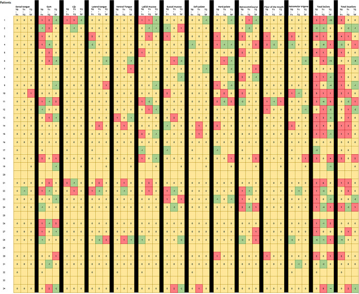
Methods: A retrospective audit of 34 patients diagnosed with proliferative verrucous leukoplakia from a university-based unit, during the period from 1995 to 2019 was performed. The mean number of visits was 23 ± 18.6. The follow-up was divided into four-time intervals to evaluate the clinical presentation, number of lesions, dysplasia grade, and malignant transformation rate.
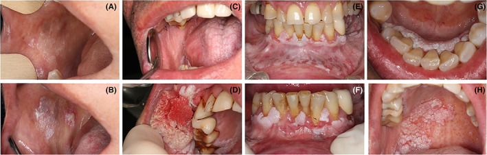
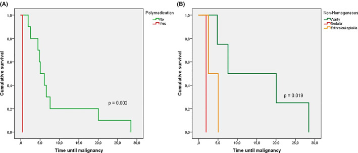
Results: The majority of patients were females 29 (85.3%), with verrucous component (77.8%), with a gingival presentation (31.8%), and with a preceding lichenoid area (44.1%). Eleven patients (32.4%) were affected by oral cancer during the follow-up, developing a total of 15 carcinomas. The mean age of malignant transformation was 67.2 ± 12.9 years, particularly 8 ± 8.5 from the onset of the lesions. Warty forms presented a higher mean estimate for malignant transformation (15.2 years, 95% confidence interval 4.4-26 years) than nodular forms (1.9 years, 95% confidence interval 1.9-1.9) (p = 0.019). Patients with an initial proliferative verrucous leukoplakia diagnosis suffered a higher risk of malignancy, particularly 15.55 times (95% confidence interval 1.69-143.17; p = 0.015) than those who did present a preceding area with lichenoid morphology.
Conclusion: Proliferative verrucous leukoplakia presented a high malignant transformation rate and sometimes displayed preceding oral lichenoid areas in early stages. Further studies are needed to understand the impact of these lichenoid areas in proliferative verrucous leukoplakia progression.
Keywords: dysplasia; malignant transformation; oral lichen planus; oral lichenoid lesions; proliferative verrucous leukoplakia.
© 2022 The Authors. Journal of Oral Pathology & Medicine published by John Wiley & Sons Ltd.
Conflict of interest statement
The authors declare no conflict of interest.
Figures
References
-
- Warnakulasuriya S. White, red, and mixed lesions of oral mucosa: a clinicopathologic approach to diagnosis. Periodontol 2000. 2019;80:89‐104. - PubMed
-
- Arduino PG, Bagan J, El‐Naggar AK, Carrozzo M. Urban legends series: oral leukoplakia. Oral Dis. 2013;19:642‐659. - PubMed
-
- Warnakulasuriya S, Kujan O, Aguirre‐Urizar JM, et al. Oral potentially malignant disorders: a consensus report from an international seminar on nomenclature and classification, convened by the WHO Collaborating Centre for Oral Cancer. Oral Dis. 2021;27:1862‐1880. - PubMed
-
- Hansen LS, Olson JA, Silverman S. Proliferative verrucous leukoplakia. A long‐term study of thirty patients. Oral Surg Oral Med Oral Pathol. 1985;60:285‐298. - PubMed
MeSH terms
LinkOut - more resources
Full Text Sources
Medical
