. 2022 Aug;9(22):e2201254.
doi: 10.1002/advs.202201254. Epub 2022 May 21.

Micro-Gel Ensembles for Accelerated Healing of Chronic Wound via pH Regulation

Affiliations

Micro-Gel Ensembles for Accelerated Healing of Chronic Wound via pH Regulation

Tingting Cui et al. Adv Sci (Weinh). 2022 Aug.

Abstract

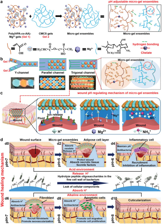
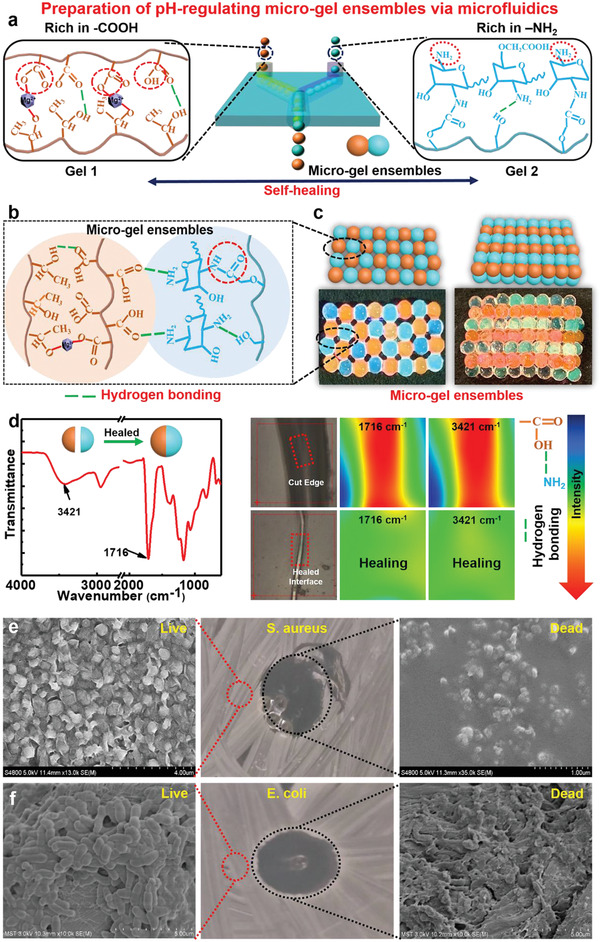
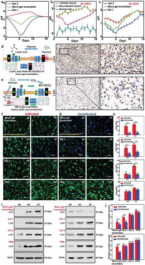
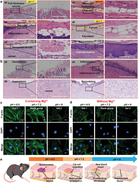
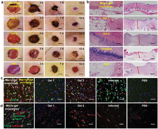
The pH value in the wound milieu plays a key role in cellular processes and cell cycle processes involved in the process of wound healing. Here, a microfluidic assembly technique is employed to fabricate micro-gel ensembles that can precisely tune the pH value of wound surface and accelerate wound healing. The micro-gel ensembles consist of poly (hydroxypropyl acrylate-co-acrylic acid)-magnesium ions (poly-(HPA-co-AA)-Mg2+ ) gel and carboxymethyl chitosan (CMCS) gel, which can release and absorb hydrogen ion (H+ ) separately at different stages of healing in response to the evolution of wound microenvironment. By regulating the wound pH to affect the proliferation and migration of cell on the wound and the activity of various biological factors in the wound, the physiological processes are greatly facilitated which results in much accelerated healing of chronic wound. This work presents an effective strategy in designing wound healing materials with vast potentials for chronic wound management.

Keywords: accelerated wound healing; chronic wound; micro-gel ensembles; microfluidic assembly; wound pH regulation.


Conflict of interest statement

The authors declare no conflict of interest.

Figures

Figure 1
Figure 1
Formation and application of micro‐gel ensembles. a) Schematic synthesis of micro‐gel ensembles via hydrogen bonding of Gel 1 and Gel 2. b) Microfluidic assembly of Gel 1 and 2 into micro‐gel ensembles with various macrostructures using specific microfluidic chips and channels. c) The pH regulating mechanism of micro‐gel ensembles for skin wound treatments, and d) the corresponding skin healing mechanism. Initially, the ‐COOH group on the surface of Gel 1 releases free H+ into the wound microenvironment, which adjust the pH of the wound during the early stage of skin healing. Then, the rich ‐NH2 group in Gel 2 absorbs free H+ from the microenvironment, converting to NH3 + and destroying the bacterial membrane structures, thus protecting the wound whilst regulating the pH value of the wound during the late stages of skin healing. Finally, micro‐gel ensembles regulate the wound's microenvironment, facilitating rapid healing process (anti‐infective, adipocyte covering wound, and macrophage polarization).
Figure 2
Figure 2
Preparation and characteristics of micro‐gel ensembles. a) Schematic illustration of preparing of micro‐gel ensembles via microfluidic technique. b) Schematic illustration of the molecular structure of micro‐gel ensembles. c) Construction of planar and 3D ordered assemblies by using Gel 1 and Gel 2 microbeads as building blocks via microfluidic assembly technique. d) IR spectrum, optical images, of Gel 1 and Gel 2 before and after self‐healing. SEM image of live/dead bacterial survival assay of e) E. coli, and f) S. aureus after in contact with the micro‐gel ensembles. n = 5 for E. coli and S. aureus.
Figure 3
Figure 3
pH regulation of micro‐gel ensembles. a) The curves of the in vivo pH values of infected wounds, non‐infected wounds, and normal skin wounds with healing time. b) The curves of the in vitro pH values of Gel 1, micro‐gel ensembles, and Gel 2 with time. Bars represent standard error, n = 5. c) The curves of the in vivo wound pH values of Gel 1, micro‐gel ensembles, and Gel 2 with healing time. Bars represent standard error, n = 5. d) Schematic illustration of lactic acid affects pH regulation, which due to insufficient oxygen supply. e) The principle of micro‐gel ensembles to reduce lactic acid and achieve accurate pH control. Scale bars of (d) and (e) are 50 µm. f) The lactic acid (green) fluorescence staining of the granulation tissue on the third and seventh day of infected S. aureus. g) The above experimental results of uninfected group. Scale bars of (f) and (g) are 20 µm. n = 5 per group. h) The changes of lactate dehydrogenase (LDH) content at different times. Data were shown as mean values ± SD. Bars represent standard error, n = 5 per group. *p < 0.05. i) The expression levels of granulation tissue in different groups as detected by Western blot analysis (WB). j) WB gray values of granulation tissue after treating wounds with different materials. Data were shown as mean values ± SD. Bars represent standard error, n = 5 per group. *p < 0.05.
Figure 4
Figure 4
In vitro and in vivo validation of adipocytes activation and fibroblast proliferation. a) H&E fluorescence staining images of new tissue at different stages after micro‐gel ensembles treatment of full‐thickness skin defects. b) H&E fluorescence staining images of the four healing stages (hemostasis, inflammation, hyperpiasia, remolding) of chronic wounds over time. The scale bars of (a) and (b) are 200 µm, n = 5 per group. Fluorescence staining images of gel materials c) with Mg2+ and d) without Mg2+ where the results showed that gel materials containing Mg2+ effectively activated the F‐actin of adipocytes in an acidic environment. The scale bars of (c) and (d) are 5 µm, n = 24. e) Schematic illustration of wound healing process (early protection, macrophage polarization, new blood formation, and tissue regeneration).
Figure 5
Figure 5
Four processes (wound healing area, granulation growth thickness, macrophage polarization, and neovascularization regeneration) of skin formation. a) Digital photographs of representative skin wound healing processes in rats treated with micro‐gel ensembles, Gel 1, Gel 2, infected, and PBS, respectively. The scale bars are 1 cm, n = 7 per group. b) Microscopy images of granulation growth of micro‐gel ensembles, Gel 1, Gel 2, infected, and PBS groups by H&E. The scale bars are 200 µm, n = 7 per group. Double immunofluorescence staining images of c) macrophages, and d) new blood vessels in the micro‐gel ensembles, Gel 1, Gel 2, infected, and PBS groups. The scale bars of (c) and (d) are 50 µm, n = 7 per group.

Similar articles

Cited by

References

    1. Hu M. S., Maan Z. N., Wu J.‐C., Rennert R. C., Hong W. X., Lai T. S., Cheung A. T., Walmsley G. G., Chung M. T., McArdle A., Longaker M. T., Lorenz H. P., Ann. Biomed. Eng. 2014, 42, 1494. - PMC - PubMed
    1. Gurtner G. C., Werner S., Barrandon Y., Longaker M. T., Nature 2008, 453, 314. - PubMed
    1. Mao C., Xiang Y., Liu X., Cui Z., Yang X., Li Z., Zhu S., Zheng Y., Yeung K. W. K., Wu S., ACS Nano 2018, 12, 1747. - PubMed
    1. Martin P., Science 1997, 276, 75. - PubMed
    1. Tan Q.‐W., Tang S.‐L., Zhang Y., Yang J.‐Q., Wang Z.‐L., Xie H.‐Q., Lv Q., J. Invest. Dermatol. 2019, 139, 455. - PubMed

Publication types

LinkOut - more resources