Review
. 2022 Nov 25;43(6):1074-1096.
doi: 10.1210/endrev/bnac013.

Hippo Signaling in the Ovary: Emerging Roles in Development, Fertility, and Disease

Affiliations
Review

Hippo Signaling in the Ovary: Emerging Roles in Development, Fertility, and Disease

Kendra L Clark et al. Endocr Rev. .

Abstract

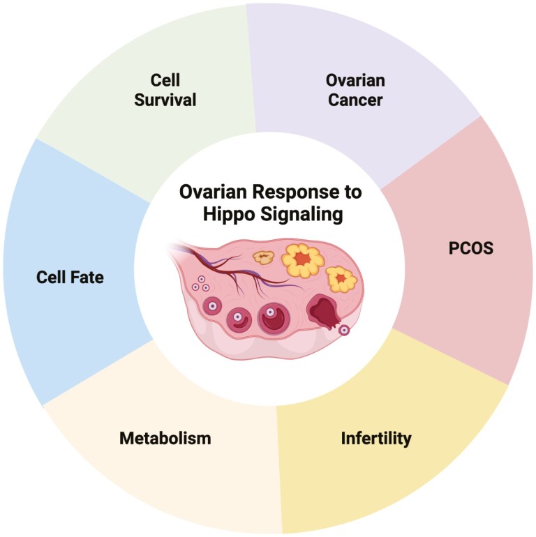
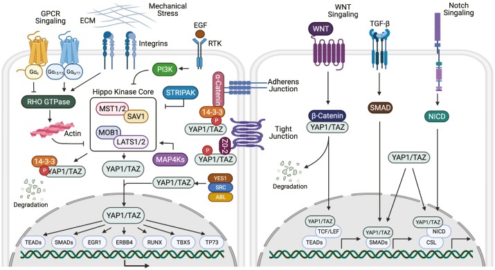
Emerging studies indicate that the Hippo pathway, a highly conserved pathway that regulates organ size control, plays an important role in governing ovarian physiology, fertility, and pathology. Specific to the ovary, the spatiotemporal expression of the major components of the Hippo signaling cascade are observed throughout the reproductive lifespan. Observations from multiple species begin to elucidate the functional diversity and molecular mechanisms of Hippo signaling in the ovary in addition to the identification of interactions with other signaling pathways and responses to various external stimuli. Hippo pathway components play important roles in follicle growth and activation, as well as steroidogenesis, by regulating several key biological processes through mechanisms of cell proliferation, migration, differentiation, and cell fate determination. Given the importance of these processes, dysregulation of the Hippo pathway contributes to loss of follicular homeostasis and reproductive disorders such as polycystic ovary syndrome (PCOS), premature ovarian insufficiency, and ovarian cancers. This review highlights what is currently known about the Hippo pathway core components in ovarian physiology, including ovarian development, follicle development, and oocyte maturation, while identifying areas for future research to better understand Hippo signaling as a multifunctional pathway in reproductive health and biology.

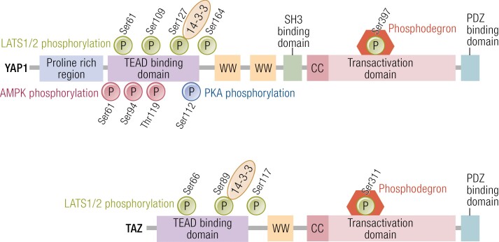
Keywords: Hippo signaling; LATS1/2; YAP1; follicle; oocyte; ovarian cancer; ovary.


Figures

Graphical Abstract
Graphical Abstract
Figure 1.
Figure 1.
Diagrammatic representation of the Hippo signaling pathway and mechanism of action in mammalian cells. The Hippo signaling pathway has many upstream regulators, from left to right: G protein–coupled receptors (GPCRs), extracellular matrix (ECM) stiffness and mechanical stress, integrins, and growth factor receptor tyrosine kinases (RTK). When core Hippo components MST1/MST2 and LATS1/LATS2 and their respective regulatory proteins SAV1 and MOB1 are phosphorylated, the Hippo pathway is active, suppressing the transcriptional activity of YAP1/TAZ by phosphorylation and sequestering YAP1/TAZ to the cytoplasm where they bind to 14-3-3 proteins or undergo protein degradation. When the Hippo pathway is inactive, dephosphorylated YAP1/TAZ enter the nucleus and bind with TEA domain transcription factors, TEADs. YAP1 and TAZ can also interact with other nuclear transcription factors. Additionally, other signaling pathways (WNT, TGF-β, and Notch) may interact with YAP1 and TAZ in order to regulate target gene expression. Abbreviations: EGF, epidermal growth factor; G, guanine nucleotide binding protein; LATS1/2, large tumor suppressor 1 and 2; MAP4K, mitogen-activated protein kinase kinase kinase kinase; MOB1, Mps one binder 1; MST1/2, mammalian STE20-like; PI3K, phosphoinositide 3-kinase; SAV1, Salvador family WW domain-containing protein 1; STRIPAK, striatin-interacting phosphatase and kinase; TAZ, transcriptional coactivator with PTZ-binding motif also called WW domain-containing transcription regulator 1, WWTR1; TGF, transforming growth factor; YAP1, yes-associated protein 1, ZO-2, zona occludens-2.
Figure 2.
Figure 2.
Structural motifs and phosphorylation sites in YAP1/TAZ. YAP1 is phosphorylated by LATS1/2 at 5 serine residues (Ser61, Ser109, Ser127, Ser164, and Ser397) and TAZ at 4 serine residues (Ser66, Ser89, Ser117, and Ser311) (green circles). Phosphorylation of YAP1 Ser127 results in the restriction of YAP1 to the cytoplasm due to binding with 14-3-3 and phosphorylation at Ser397 activates a phosphodegron degradation motif leading to reduced levels of YAP1 and TAZ. Likewise, phosphorylation of TAZ Ser89 sequesters TAZ to the cytoplasm via binding with 14-3-3 and Ser 311 activates a phosphodegron degradation motif. Phosphorylation of YAP1 by AMP-activated protein kinase (AMPK) occurs at multiple sites shown by red circles. Phosphorylation at Ser 94 via AMPK disrupts the YAP-TEAD interactions, resulting in reduction of the transcription and YAP1 target genes. Protein kinase A (PKA) activates LATS and results in LATS mediated phosphorylation of YAP1 and TAZ. Phosphorylation of YAP1 at Ser 112 by PKA enables YAP1 to translocate to the nucleus (blue circle). Abbreviations: cc, coiled-coil domain; ww, WW domain.

Similar articles

Cited by

References

    1. Matsuda F, Inoue N, Manabe N, Ohkura S. Follicular growth and atresia in mammalian ovaries: regulation by survival and death of granulosa cells. J Reprod Dev. 2012;58(1):44-50. - PubMed
    1. Vanselow J, Christenson LK, Pate JL. Editorial: regulation of dynamic changes and remodeling events during the formation, rescue and regression of the corpus luteum. Front Endocrinol. 2020;11:244. - PMC - PubMed
    1. Brown HM, Russell DL. Blood and lymphatic vasculature in the ovary: development, function and disease. Hum Reprod Update. 2014;20(1):29-39. - PubMed
    1. Ishizuka B. Current understanding of the etiology, symptomatology, and treatment options in premature ovarian insufficiency (POI). Front Endocrinol. 2021;12:626924. - PMC - PubMed
    1. Hirshfield AN. Development of follicles in the mammalian ovary. Int Rev Cytol. 1991;124:43-101. - PubMed

Publication types