

Early root migration after a mandibular third molar coronectomy
- PMID: 35596808
- PMCID: PMC9123869
- DOI: 10.1007/s10006-022-01072-z
Early root migration after a mandibular third molar coronectomy
Erratum in
-
Correction to: Early root migration after a mandibular third molar coronectomy.Oral Maxillofac Surg. 2023 Sep;27(3):545. doi: 10.1007/s10006-022-01079-6. Oral Maxillofac Surg. 2023. PMID: 35657575 Free PMC article. No abstract available.
Abstract
Purpose: This prospective cohort study aimed to assess early root migration after a coronectomy of the mandibular third molar at 2 and 6 months after surgery.
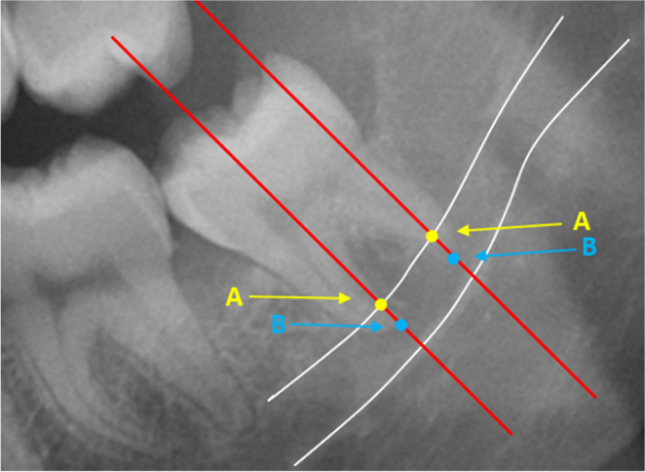
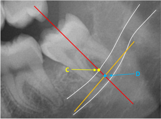
Methods: We included all patients treated with a coronectomy of an impacted mandibular third molar. The primary outcome measure was the extent of postoperative root migration after 2 and 6 months. Migration was measured as the distance between the root complex and a fixed point on the inferior alveolar canal. The secondary aim was to identify factors (age, impaction pattern, and patient sex) that affected the extent of root migration.
Results: One hundred and sixty-five coronectomies were performed in 141 patients (96 females and 45 males; mean age 33.1 years, SD 16.0). The 2-month checkup was completed by 121 patients that received 141 coronectomies. The 6-month check-up was completed by 73 patients that received 80 coronectomies. The mean root migrations were 3.30 mm (SD 2.53 mm) at 2 months and 5.27 mm (SD 3.14 mm) at 6 months. In the 2-6-month interval, the mean root migration was 2.58 mm (SD 2.07 mm). The extents of migration were similar during the 0-2-month interval and the 2-6-month interval (p = 0.529). Younger age was associated with greater root migration, and females experienced significantly greater migrations than males (p = 0.002).
Conclusion: Roots migrated more rapidly in the first two postoperative months, compared to the 2-6-month interval. Age was negatively correlated with the extent of root migration, and females showed significantly greater migrations than males.
Keywords: Coronectomy; Mandibular third molar; Root migration; Third molars.
© 2022. The Author(s).
Conflict of interest statement
The authors declare no competing interests.
Figures


References
-
- Renton T, Hankins M, Sproate C, McGurk M. A randomised controlled clinical trial to compare the incidence of injury to the inferior alveolar nerve as a result of coronectomy and removal of mandibular third molars. Br J Oral Maxillofac Surg. 2005;43:7–12. doi: 10.1016/j.bjoms.2004.09.002. - DOI - PubMed
-
- Kouwenberg AJ, Stroy LPP, Vree- vd Rijt ED, Mensink G, Gooris PJJ (2016) Coronectomy of the mandibular third molar: Respect for the inferior alveolar nerve. J Cranio-Maxillofacial Surg 44: 616-621 - PubMed
MeSH terms
LinkOut - more resources
Full Text Sources
