. 2022 May 21;24(2):18.
doi: 10.1007/s10544-022-00617-z.

Well-free agglomeration and on-demand three-dimensional cell cluster formation using guided surface acoustic waves through a couplant layer

Affiliations

Well-free agglomeration and on-demand three-dimensional cell cluster formation using guided surface acoustic waves through a couplant layer

Jiyang Mei et al. Biomed Microdevices. .

Abstract

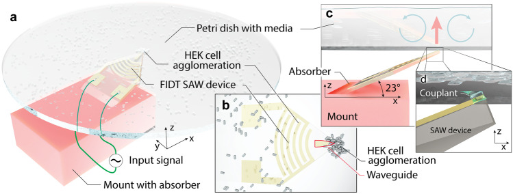
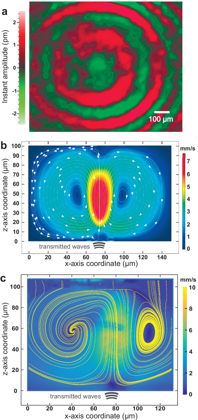
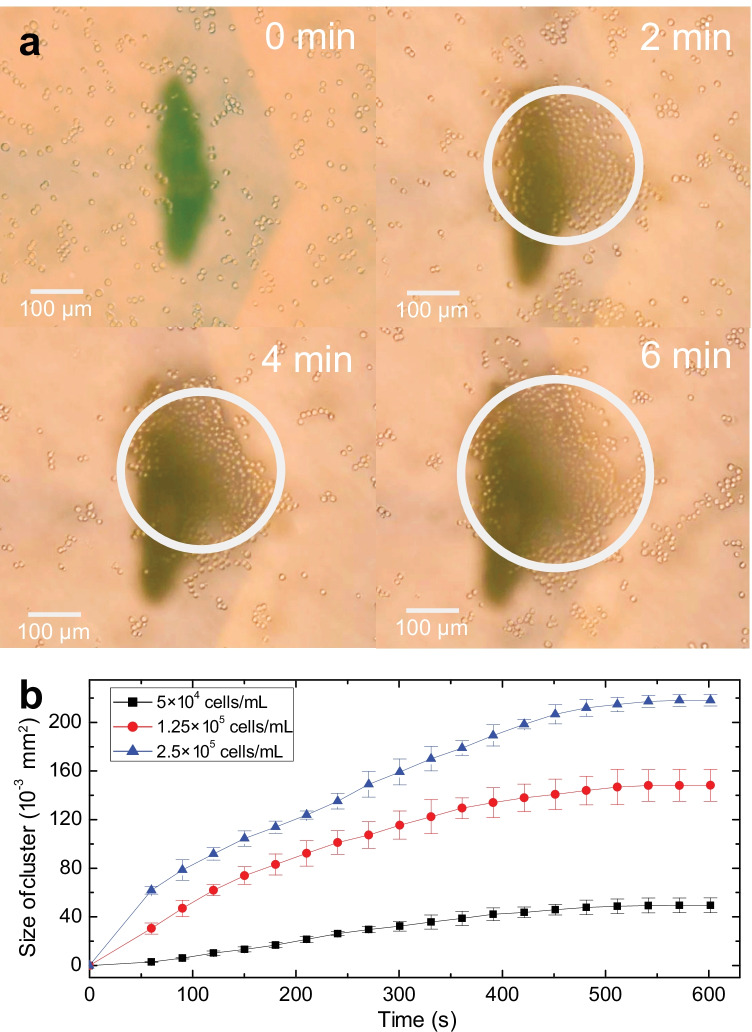
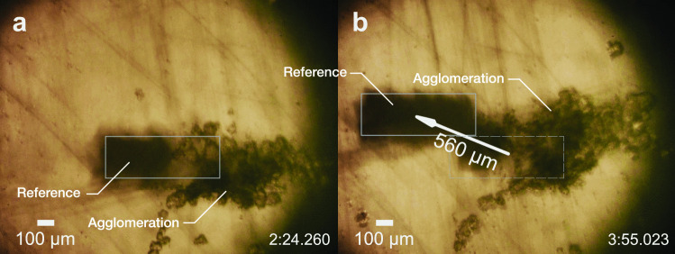
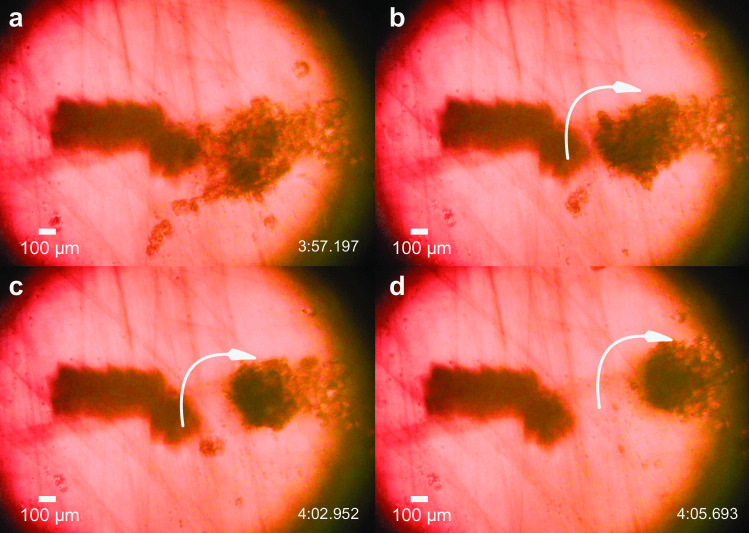
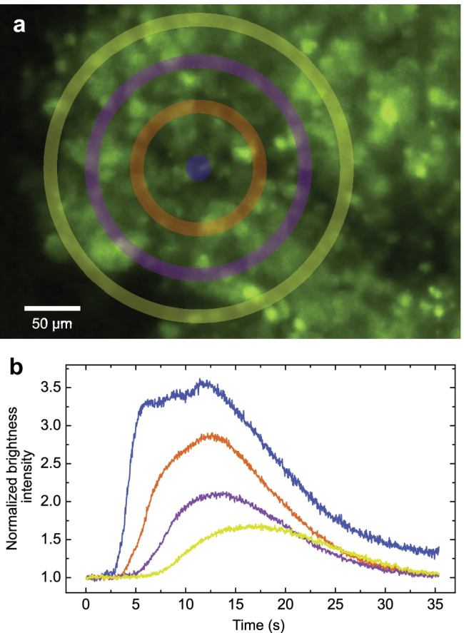
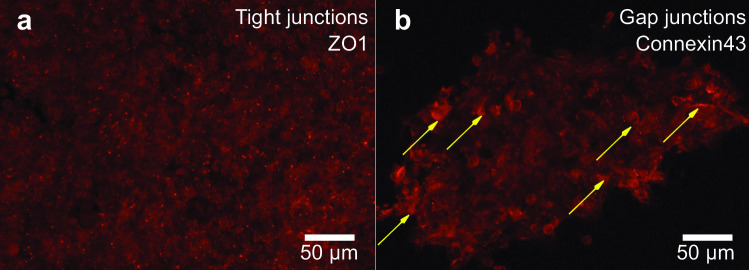
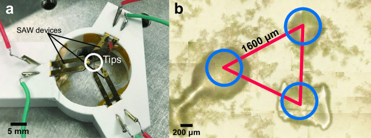
Three-dimensional cell agglomerates are broadly useful in tissue engineering and drug testing. We report a well-free method to form large (1.4-mm) multicellular clusters using 100-MHz surface acoustic waves (SAW) without direct contact with the media or cells. A fluid couplant is used to transform the SAW into acoustic streaming in the cell-laden media held in a petri dish. The couplant transmits longitudinal sound waves, forming a Lamb wave in the petri dish that, in turn, produces longitudinal sound in the media. Due to recirculation, human embryonic kidney (HEK293) cells in the dish are carried to the center of the coupling location, forming a cluster in less than 10 min. A few minutes later, these clusters may then be translated and merged to form large agglomerations, and even repeatedly folded to produce a roughly spherical shape of over 1.4 mm in diameter for incubation-without damaging the existing intercellular bonds. Calcium ion signaling through these clusters and confocal images of multiprotein junctional complexes suggest a continuous tissue construct: intercellular communication. They may be formed at will, and the method is feasibly useful for formation of numerous agglomerates in a single petri dish.

Keywords: Acoustofluidics; Cell agglomerate; Surface acoustic wave.


Conflict of interest statement

There are no competing interests to declare.

Figures

Fig. 1
Fig. 1
The cell agglomeration device. The a lithium niobate (LN) substrate is held in place by a mount with an absorber to prevent spurious and reflected acoustic waves at the Rayleigh angle of 23. This structure is mounted below a petri dish laden with media and HEK cells. Focusing surface acoustic waves generated by an input signal into a (b) focusing interdigital transducer (FIDT) on a lithium niobate substrate propagate into the superstrate (petri dish) through an Au focusing waveguide (thickness exaggerated, outlined in red) and into <0.2 μL Tween 20 as a couplant. The b view is from above and along the -z axis through the media, cells, and petri dish. The mount is omitted for clarity. Acoustic energy is passed c vertically through the d couplant (along the z axis), through the petri dish, and into the cell-laden media (thick arrow). Acoustic streaming is generated in the media, in turn leading to a (c) local recirculation region (thin arrows) around the coupling position. This, in turn, leads to (a, b) cell agglomeration above the SAW device’s 40 μm wide tip. (Cells, Au thickness not to scale for clarity)
Fig. 2
Fig. 2
Generation of local vortical flow by leaky Lamb waves from the coupling point. a LDV measurements display the Lamb wave propagating along the petri dish, from roughly at the center, the source of acoustic energy from coupling with the SAW device from below. The Lamb wave concentrically spreads out from the center of transmission, where red and green colors denote the instantaneous peaks and valleys of the vibration. Scale bar: 100 μm. Side views of the recirculation in the fluid within the petri dish, as actuated by the transmitted acoustic waves from the SAW device through the couplant liquid, and onward through the glass of the petri dish into the fluid within, using b finite element analysis, and c experimental μPIV, where the background color represents the velocity magnitude and the yellow lines display the streamlines. The petri dish’s top surface is at z=0, and the couplant is centered at x=78μm along the x axis
Fig. 3
Fig. 3
Cell agglomeration via coupled SAW-Lamb waves. Initially, cells are homogeneously distributed in the media at a concentration of 1.25×10⁵ cells/mL with no SAW. Upon activating 92 mW SAW in this arrangement, cells are gradually accumulated above the coupling point where the acoustic wave is transmitted from below, indicated in the images above with a dark right-handed chevron shape. The white circles outline the grouping of cells that gradually grow over time until they reach an almost steady state. This is more clearly shown by b) a plot of the cluster’s area with respect to time at various cell concentrations, 5×10⁴ cells/mL (black squares), 1.25×10⁵ cells/mL (red circles), 2.5×10⁵ cells/mL (blue triangles). After eight minutes, the cluster size reached a steady state in this experiment. The error bars denote the standard deviation from five measurements of the cluster size. A video of the phenomenon is provided in the Supplementary Information. Scale bar: 100 μm
Fig. 4
Fig. 4
Translation of cell agglomerations in the petri dish. After agglomeration and waiting (with the transducer off) for about five minutes, the cell cluster may be transported along with the tip of the transducer underneath the petri dish. The (boxed) reference mark as shown is attached to the petri dish. By leaving the transducer coupling tip and the observation microscope fixed in place, and translating the dish up and to the left by 560 μm (for example) over a period of 90 s—from a 2 min 24.260 s to b 3 min 55.023 s—the agglomeration was moved downward and to the right by this distance relative to the petri dish. Scale bar: 100 μm
Fig. 5
Fig. 5
Cell agglomerate folding and rolling. After waiting for 5 min to weakly bind the existing cells together, increasing the input power to 350 mW causes the cluster to (a, b) roll upon itself from the left edge, folding atop the remainder of the cells and forming (c,d) a roughly spherical cell agglomeration in 8 s. Scale bar: 100 μm. Supplementary Video 2 shows the process in real time
Fig. 6
Fig. 6
Calcium ion signaling in a cell agglomerate after 22 h incubation. The calcium (in green) transmission in a 3D cell agglomerate in the order from center to periphery (blue-orange-magenta-yellow). a The concentric regions of interest denoted on a photo of the cluster acquired from fluorescent microscopy. b The normalized brightness intensity mapping across of the corresponding regions. The Ca2+ signaling seen here and in the video in the Supplementary Information indicates the cells are functioning as a collective group. Scale bar: 50 μm
Fig. 7
Fig. 7
Indication of tight and gap junctions in the cell agglomeration. Confocal images of multiprotein junctional complexes (in red) in acoustically-formed 3D clusters. a Tight junctions in the cluster support intracellular bonding, maintaining the aggregated structure. b Gap junctions (stronger signals marked by yellow arrows) permit intercellular communication, including Ca2+ propagation in the cluster as shown in Fig. 6. Scale bar: 50 μm
Fig. 8
Fig. 8
Simultaneous formation of multiple agglomerates. a The platform holding three mounted SAW devices could transmit waves to the superstrate at three distinct locations. b Three clusters, at sizes of about 500 μm, were simultaneously made in the petri dish

Similar articles

Cited by

References

    1. A. Abbott, Biology’s new dimension. Nature Publishing Group (2003) - PubMed
    1. D.R. Albrecht, G.H. Underhill, T.B. Wassermann, R.L. Sah, S.N. Bhatia, Probing the role of multicellular organization in three-dimensional microenvironments. Nat. Methods. 3(5), 369–375 (2006) - PubMed
    1. Bao G, Suresh S. Cell and molecular mechanics of biological materials. Nat. Mater. 2003;2(11):715–725. doi: 10.1038/nmat1001. - DOI - PubMed
    1. Bartfeld S, Bayram T, van de Wetering M, Huch M, Begthel H, Kujala P, Vries R, Peters PJ, Clevers H. In vitro expansion of human gastric epithelial stem cells and their responses to bacterial infection. Gastroenterology. 2015;148(1):126–136. doi: 10.1053/j.gastro.2014.09.042. - DOI - PMC - PubMed
    1. Bersini S, Jeon JS, Dubini G, Arrigoni C, Chung S, Charest JL, Moretti M, Kamm RD. A microfluidic 3d in vitro model for specificity of breast cancer metastasis to bone. Biomaterials. 2014;35(8):2454–2461. doi: 10.1016/j.biomaterials.2013.11.050. - DOI - PMC - PubMed

Publication types