Review
. 2022 Jul;12(7):1501-1533.
doi: 10.1007/s13555-022-00737-7. Epub 2022 May 21.

Current and Emerging Strategies to Inhibit Type 2 Inflammation in Atopic Dermatitis

Affiliations
Review

Current and Emerging Strategies to Inhibit Type 2 Inflammation in Atopic Dermatitis

El-Bdaoui Haddad et al. Dermatol Ther (Heidelb). 2022 Jul.

Abstract

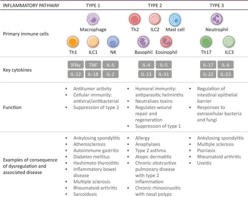
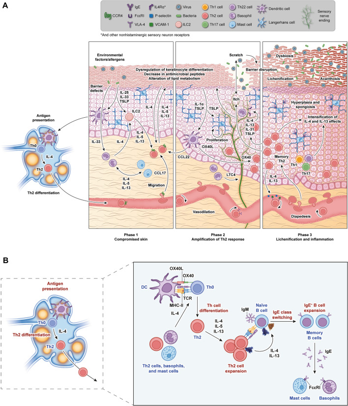
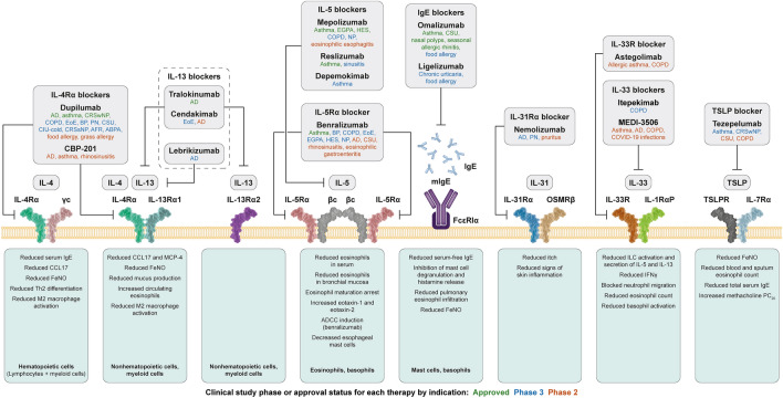
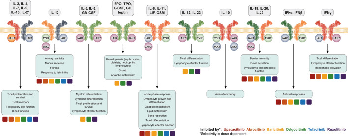
Type 2 immunity evolved to combat helminth infections by orchestrating a combined protective response of innate and adaptive immune cells and promotion of parasitic worm destruction or expulsion, wound repair, and barrier function. Aberrant type 2 immune responses are associated with allergic conditions characterized by chronic tissue inflammation, including atopic dermatitis (AD) and asthma. Signature cytokines of type 2 immunity include interleukin (IL)-4, IL-5, IL-9, IL-13, and IL-31, mainly secreted from immune cells, as well as IL-25, IL-33, and thymic stromal lymphopoietin, mainly secreted from tissue cells, particularly epithelial cells. IL-4 and IL-13 are key players mediating the prototypical type 2 response; IL-4 initiates and promotes differentiation and proliferation of naïve T-helper (Th) cells toward a Th2 cell phenotype, whereas IL-13 has a pleiotropic effect on type 2 inflammation, including, together with IL-4, decreased barrier function. Both cytokines are implicated in B-cell isotype class switching to generate immunoglobulin E, tissue fibrosis, and pruritus. IL-5, a key regulator of eosinophils, is responsible for eosinophil growth, differentiation, survival, and mobilization. In AD, IL-4, IL-13, and IL-31 are associated with sensory nerve sensitization and itch, leading to scratching that further exacerbates inflammation and barrier dysfunction. Various strategies have emerged to suppress type 2 inflammation, including biologics targeting cytokines or their receptors, and Janus kinase inhibitors that block intracellular cytokine signaling pathways. Here we review type 2 inflammation, its role in inflammatory diseases, and current and future therapies targeting type 2 pathways, with a focus on AD. INFOGRAPHIC.

Keywords: Atopic dermatitis; Biologic; Cytokine; Janus kinase; Type 2 inflammation.


Figures

Fig. 1
Fig. 1
Overview of types 1, 2, and 3 inflammation. This diagram represents immune concepts that do not occur in complete isolation. IFNγ interferon-γ, IL interleukin, ILC innate lymphoid cell, NK natural killer, Th1/2 T helper type 1/2 cell, TNF tumor necrosis factor
Fig. 2
Fig. 2
Pathologic inflammation in AD (A), and IL-4 as a key driver of Th2 differentiation and activation (B). CCL17 C–C motif chemokine ligand 17, DC dendritic cell, Ig immunoglobin, IL interleukin, ILC innate lymphoid cell, LTC4 leukotriene C4, MHC-II major histocompatibility complex II, TCR T-cell receptor, TSLP thymic stromal lymphopoietin, VCAM-1 vasopressin-activated calcium-mobilizing receptor 1
Fig. 3
Fig. 3
Biologics that inhibit type 2 molecules. AD atopic dermatitis, ADCC antibody-dependent cellular cytotoxicity, ABPA allergic bronchopulmonary aspergillosis, AFR allergic fungal rhinosinusitis, BP bullous pemphigoid, CCL17 C–C motif chemokine ligand 17, COPD chronic obstructive pulmonary disorder, CRSsNP chronic rhinosinusitis without nasal polyposis, CRSwNP chronic rhinosinusitis with nasal polyposis, CIU chronic idiopathic urticaria, CSU chronic spontaneous urticaria, EGPA eosinophilic granulomatosis with polyangiitis, EoE eosinophilic esophagitis, FeNO fractional exhaled nitric oxide, HES hypereosinophilic syndrome, IFNγ interferon-gamma, IgE immunoglobulin E, IL interleukin, MCP-4 monocyte chemoattractant protein-4, NP nasal polyps, OSMRβ oncostatin-M specific receptor subunit β, PC20 provocative concentration causing a 20% drop in FEV1 from baseline, PN prurigo nodularis, TSLP thymic stromal lymphopoietin, TSLPR thymic stromal lymphopoietin receptor
Fig. 4
Fig. 4
JAK signaling pathways in type 2 inflammation and JAK inhibitors. Adapted from Schwartz et al. [238]. For simplicity, the depicted receptor subunits do not reflect the extent of differentiation in domain structures of the various JAK cytokine receptors. EPO erythropoietin, G-CSF granulocyte colony-stimulating factor, GH growth hormone, GM-CSF granulocyte macrophage colony-stimulating factor, IFN interferon, IL interleukin, JAK Janus kinase, LIF leukemia inhibitory factor, OSM oncostatin M, TPO thrombopoietin

Similar articles

Cited by

References

    1. Chaplin DD. Overview of the immune response. J Allergy Clin Immunol. 2010;125(2 suppl 2):S3–23. - PMC - PubMed
    1. Palm NW, Rosenstein RK, Medzhitov R. Allergic host defences. Nature. 2012;484(7395):465–472. - PMC - PubMed
    1. Maizels RM. Regulation of immunity and allergy by helminth parasites. Allergy. 2020;75(3):524–534. - PubMed
    1. Inclan-Rico JM, Siracusa MC. First responders: innate immunity to helminths. Trends Parasitol. 2018;34(10):861–880. - PMC - PubMed
    1. de Kouchkovsky DA, Ghosh S, Rothlin CV. Negative regulation of type 2 immunity. Trends Immunol. 2017;38(3):154–167. - PMC - PubMed