. 2022 Jun 2;82(11):2098-2112.e4.
doi: 10.1016/j.molcel.2022.04.026. Epub 2022 May 20.

A hexasome is the preferred substrate for the INO80 chromatin remodeling complex, allowing versatility of function

Affiliations

A hexasome is the preferred substrate for the INO80 chromatin remodeling complex, allowing versatility of function

Laura J Hsieh et al. Mol Cell. .

Abstract

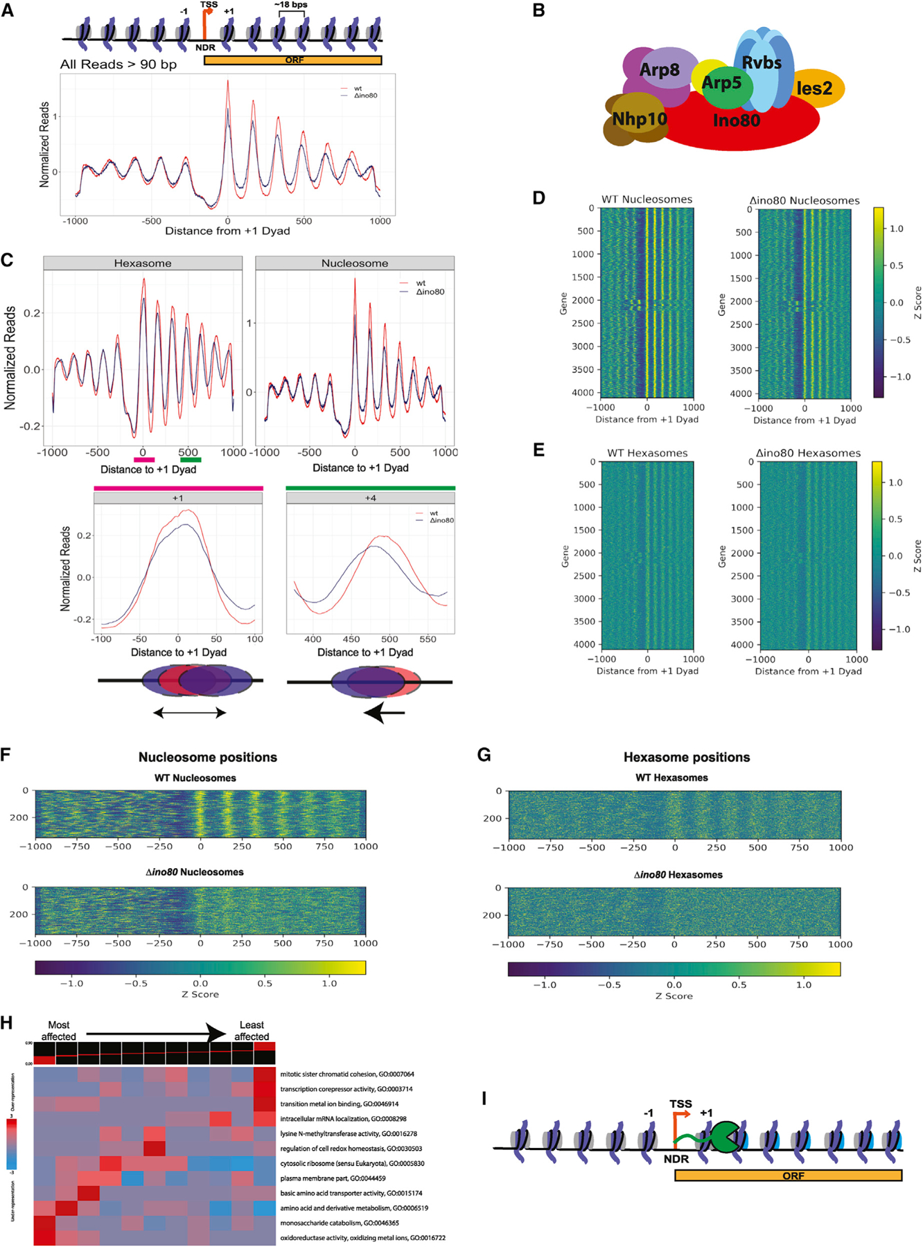
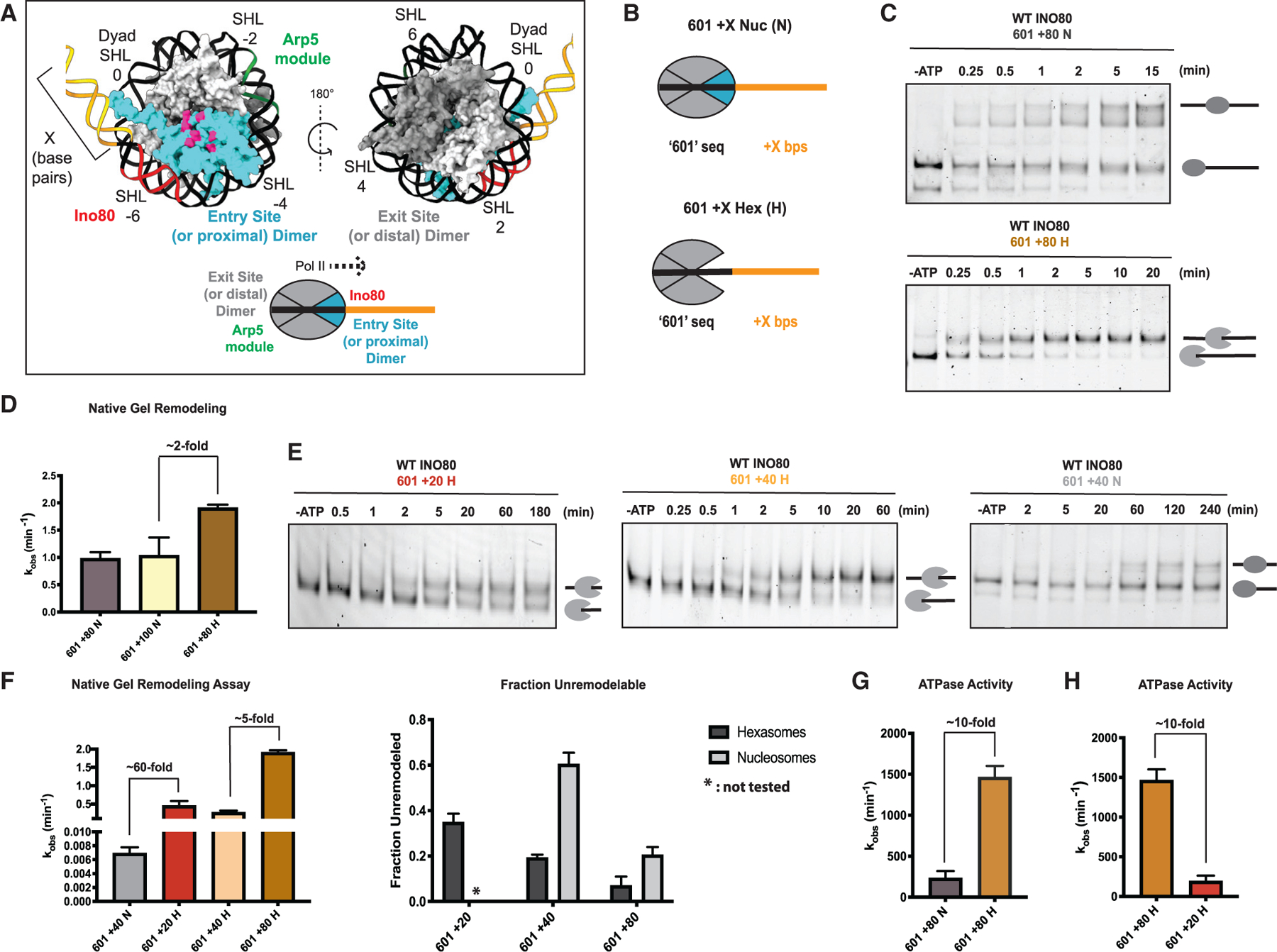
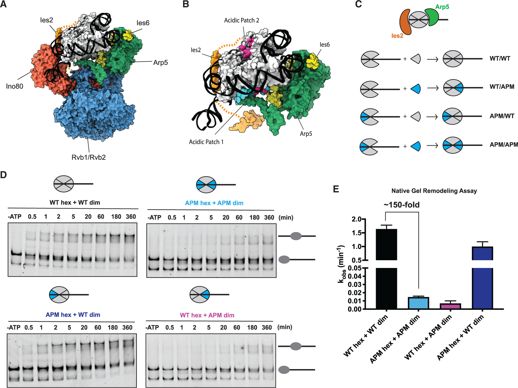
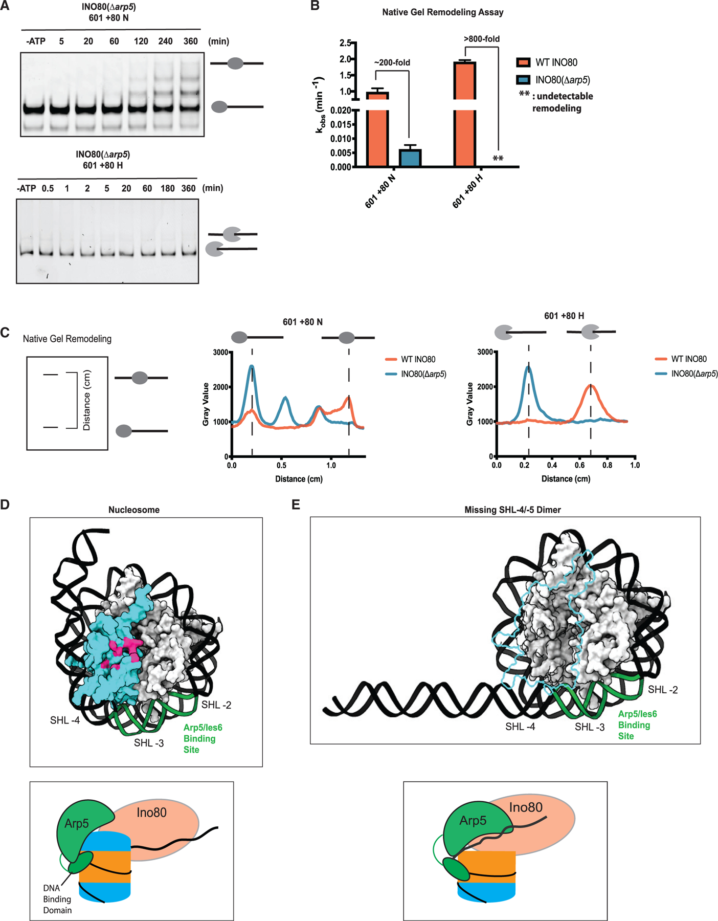
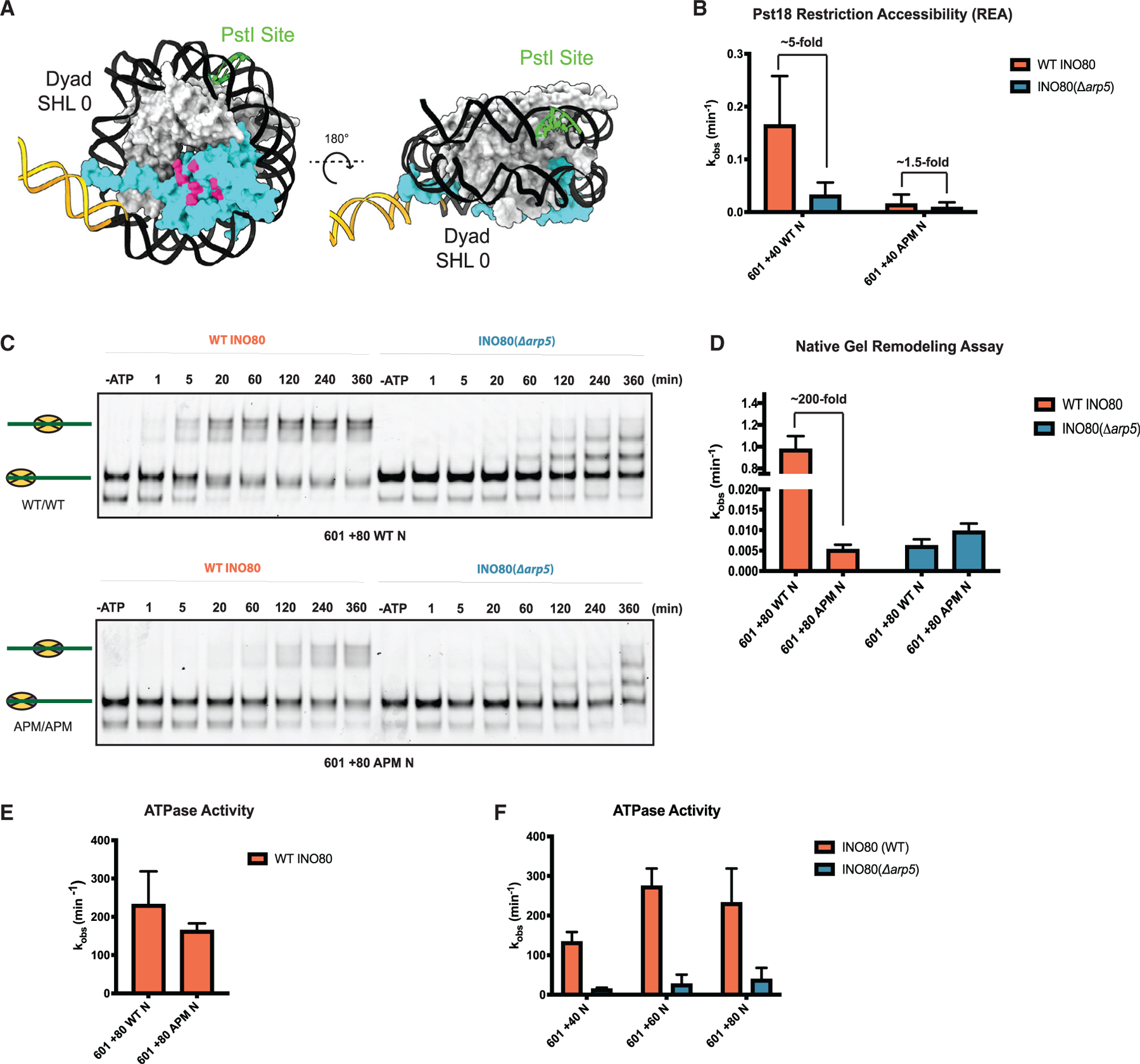
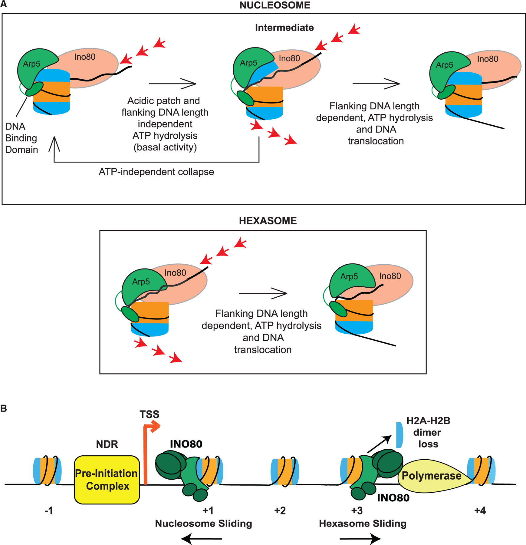
The critical role of the INO80 chromatin remodeling complex in transcription is commonly attributed to its nucleosome sliding activity. Here, we have found that INO80 prefers to mobilize hexasomes over nucleosomes. INO80's preference for hexasomes reaches up to ∼60 fold when flanking DNA overhangs approach ∼18-bp linkers in yeast gene bodies. Correspondingly, deletion of INO80 significantly affects the positions of hexasome-sized particles within yeast genes in vivo. Our results raise the possibility that INO80 promotes nucleosome sliding by dislodging an H2A-H2B dimer, thereby making a nucleosome transiently resemble a hexasome. We propose that this mechanism allows INO80 to rapidly mobilize nucleosomes at promoters and hexasomes within gene bodies. Rapid repositioning of hexasomes that are generated in the wake of transcription may mitigate spurious transcription. More generally, such versatility may explain how INO80 regulates chromatin architecture during the diverse processes of transcription, replication, and repair.

Keywords: INO80; chromatin remodeling; hexasome.


Conflict of interest statement

Declaration of interests Geeta Narlikar is on the Molecular Cell advisory board.

Figures

Figure 1.
Figure 1.. INO80 regulates positions of subnucleosomal particles in vivo
(A and B) (A) Normalized MNase signal at the TSSs of all genes for WT, in orange, and Δino80, in dark blue, for all fragment lengths greater than 90 bps. The x axis represents distance from +1 nucleosome dyad. Upper panel: schematic of the corresponding chromatin architecture in (B) illustration of the INO80 complex. Only one subunit per module is labeled. (C) Upon deletion of Ino80, nucleosomes and hexasomes are less well positioned at +1 location and display shifted positions in gene body. Upper panel: fragments were binned by sizes representing either hexasomes (100 ± 10 bps) or nucleosomes (147 ± 10 bps), and the average signal at TSSs and in the gene body are plotted for WT and Δino80. Lower panel: pink and green lines in the hexasome plot from the upper panel are magnified to show hexasome footprints at +1 and +4 positions. Below the graphs are illustrations of the respective changes in positioning that occur from WT (red) to Δino80 (blue). (D) Heatmap of nucleosome footprint signals across all genes for WT and Δino80 cells. (E) Same as (D) but for the hexasome footprint signals. (F) Data for genes where nucleosome positions are most affected by deletion of Ino80. A heatmap representing genes that had the lowest Spearman Rho correlation (i.e., most affected by deletion of Ino80) for nucleosome footprint signals ranging from the −100 to +1,000 bps of the +1 dyad between WT and Δino80. (G) Data for the genes in (F) but focusing on hexasome positions. (H) An iPAGE heatmap of the correlations of the nucleosome footprint signal between WT and Δino80 and their annotated GO terms. The genes have been binned into 11 groups, with the lowest correlation group (most affected in terms of nucleosome footprints) on the left, and the highest correlation group (least affected in terms of nucleosome footprints) on the right. (I) Illustration of RNA Pol II traversing through the gene body. The promoter distal dimer that is lost during elongation is depicted in cyan.
Figure 2.
Figure 2.. Hexasomes are better substrates for INO80 in vitro
(A) Upper panel: two different rotations of nucleosome structure (PDB: 1KX5) highlighting the dimer (cyan) at the entry site (or proximal to flanking DNA) and its acidic patch (pink). Regions of DNA where Arp5-Ies6 binds (green), Ino80 ATPase binds (red), that are in flanking DNA (yellow), and are in super helical locations (SHLs) are indicated. Lower panel: illustration of nucleosome, indicating the exit site (or distal) and entry site (or proximal) H2A-H2B dimers, and regions where the Arp5-Ies6 and Ino80 ATPase binds. Direction of elongating RNA Pol II based on the loss of the distal dimer is shown with a black dotted line. (B) Depiction of 601 nucleosome (N) and hexasome (H) constructs with flanking DNA used in this study. The H2A-H2B dimer missing in hexasomes is in cyan. (C) Example native gels showing remodeling by INO80 of 601 + 80 nucleosomes (top) and 601 + 80 hexasomes (bottom). Substrates (end-positioned nucleosomes or end-positioned hexasomes) are labeled by illustrations next to the respective bands in gels. (D) Quantification of rate constants from multiple repeats of data, such as in (C). Rate constant for remodeling of 601 + 100 N is also shown. (E) Example gels of INO80 remodeling with 601 + 20 H (left), 601 + 40 H (middle), and 601 + 40 N (right). Substrates (end-positioned nucleosomes or end-positioned hexasomes) are labeled by illustrations next to the respective bands in gels. (F) Left panel: quantification of rate constants for remodeling of 601 + 40 N (gray), 601 +20 H (red), 601 + 40 H (peach), and 601 + 80 H (tan) by INO80 as assayed using the native gel assay. Note that the data for 601 + 80 H are the same data shown in (D) and are shown again here for ease of comparison. Right panel: fraction of unremodeled substrate, assayed via the native gel assay and measured at the longest time point (where all reactions have mostly gone to completion). Hexasomes are in dark gray, and nucleosomes are in light gray. (G) Observed rate constants of INO80 ATPase activity on 601 + 80 N in gray and 601 + 80 H in tan. ATPase assays were performed under the same conditions as native-gel-based remodeling. (H) Rate constants of INO80 ATPase activity on 601 + 80 H (tan) (data from G) and 601 + 20 H (red). Note that the data for 601 + 80 H are the same data as shown in (G) and are shown again here for ease of comparison. Error bars represent SEM from n > 3.
Figure 3.
Figure 3.. The acidic patch that binds to Ies2 is dispensable for INO80 sliding
(A) Cryo-EM-based structure of the core INO80 components bound to the nucleosome (PDB: 6FML). (B) Cryo-EM model from (A) showing only the Arp5 (green)-Ies6 (yellow) module and Ies2 (orange) bound to the nucleosome for clarity. Arp5-Ies6 and Ies2 bind to acidic patches (pink) on opposite sides of the nucleosome (PDB: 6FML). (C) Schematic showing assembly of nucleosomes, with acidic patch mutations (APMs) depicted in blue. From (A), Arp5 module (green) binds dimer at the entry site, whereas Ies2 (orange) binds dimer at the exit site. (D) Example native gels showing remodeling time courses for nucleosomes shown (C). (E) Quantification of rate constants from multiple repeats of data, such as that shown in (D). Error bars represent SEM from n > 3.
Figure 4.
Figure 4.. The Arp5 module is a key regulatory component for remodeling
(A) Example time courses for remodeling of 601 + 80 N (top panel) and 601 + 80 H (bottom panel) by INO80(Δarp5) as assayed by native gel. (B) Quantification of INO80(Δarp5) remodeling rate constants (blue) on 601 + 80 N and 601 + 80 H from repeats of data, such as in (A). For ease of comparison, the same WT INO80 data shown in Figure 2D are shown again here (coral). (C) Left panel: schematic showing the distance parameter in the line-scan data depicted in the middle and right panels. Middle panel: line scan showing distribution of band intensity (gray value) on a native gel for the last time point for remodeling of 601 + 80 N by WT INO80 (coral, 360 min) and INO80(Δarp5) (blue, 360 min). Right panel: line scan showing the distribution of band intensity (gray value) on a native gel for the last time point for remodeling of 601 + 80 H by WT INO80 (coral, 360 min) and INO80(Δarp5) (blue, 360 min). (D) Top panel: cryo-EM structure of the nucleosome (PDB: 6FML) showing the entry site dimer (cyan) and the Arp5 module’s DNA-binding site (green) at SHL-2/-3 (from PDB: 6FML). Lower panel: illustration of the Arp5 module binding the nucleosome (H2A-H2B dimers in cyan; H3-H4 tetramer in orange; DNA in black) at the designated binding site via a DNA-binding domain, based on PDB: 6FML. (E) Top panel: model of the structure of a hexasome missing the entry site dimer. Model shows how the wrapping of the DNA may change upon proximal dimer loss to make Arp5 module’s binding site at SHL-2/-3 (in green) more accessible. Lower panel: illustrated model of the Arp5 module binding the hexasome in a different conformation due to the absence of the dimer. Error bars represent SEM from n > 3.
Figure 5.
Figure 5.. The Arp5 module interacts with the acidic patch to regulate INO80 sliding
(A) Model of the nucleosome (PDB: 1KX5) from different angles with location of the engineered PstI restriction site in green. Proximal H2A-H2B dimer is in cyan. (B) Rate constants of cutting by PstI for WT and double APM 601 + 40 N, with saturating ATP and saturating WT INO80 (coral) or INO80(Δarp5) (blue). (C) Example native gels showing remodeling of WT (top panel) and double APM (bottom panel) 601 + 80 N with WT INO80 (coral) and INO80(Δarp5) (blue). (D) Quantification of rate constants from multiple repeats of data, such as that shown in (C). (E) Rate constants for INO80 ATPase activity with WT and double APM, 601 + 80 N. All ATPase assays were performed under the same conditions as native gel remodeling. (F) Observed rate constants for ATPase activity of WT INO80 (coral) and INO80(Δarp5) (blue) on 601 + 40 N, 601 + 60 N, and 601 + 80 N. Error bars represent SEM from n > 3.
Figure 6.
Figure 6.. Model of the INO80 remodeling mechanism
(A) Illustration of the model for INO80 action on a nucleosome and a hexasome. Nucleosome binding stimulates a basal level of ATP hydrolysis and the Ino80 motor pumps DNA into the nucleosome. This ATPase activity is independent of flanking DNA length and the acidic patch. The torsional strain caused by the pumping of the DNA is partially relieved through transient dislodging of the H2A-H2B dimer. Such dislodging is speculated to occur by the Arp5 module through contacts with the acidic patch and DNA at SHL-2/-3. This transition results in the formation of an intermediate, which can either collapse back in an ATP-independent manner, or transition forward in an ATP-dependent manner to translocate DNA across the nucleosome. Translocation is dictated by flanking DNA length and requires flanking DNA-length-dependent ATP hydrolysis. In comparison, for a hexasome, because the dimer is absent and does not inhibit INO80 remodeling, translocation occurs more readily. (B) Schematic of INO80 participating in chromatin remodeling at sites of transcription. At the +1 location, INO80 helps position the nucleosome. During elongation as RNA Pol II actively removes the H2A-H2B dimer distal to the promoter, INO80 can act on these subnucleosomal particles to restore proper positioning and help prevent aberrant transcription initiation.

References

    1. Alcid EA, and Tsukiyama T (2014). ATP-dependent chromatin remodeling shapes the long noncoding RNA landscape. Genes Dev 28, 2348–2360. - PMC - PubMed
    1. Ayala R, Willhoft O, Aramayo RJ, Wilkinson M, McCormack EA, Ocloo L, Wigley DB, and Zhang X (2018). Structure and regulation of the human INO80-nucleosome complex. Nature 556, 391–395. - PMC - PubMed
    1. Belotserkovskaya R, Oh S, Bondarenko VA, Orphanides G, Studitsky VM, and Reinberg D (2003). FACT facilitates transcription-dependent nucleosome alteration. Science 301, 1090–1093. - PubMed
    1. Brahma S, Udugama MI, Kim J, Hada A, Bhardwaj SK, Hailu SG, Lee T-H, and Bartholomew B (2017). INO80 exchanges H2a.Z for H2A by translocating on DNA proximal to histone dimers. Nat. Commun 8, 15616. - PMC - PubMed
    1. Brahma S, Ngubo M, Paul S, Udugama M, and Bartholomew B (2018). The Arp8 and Arp4 module acts as a DNA sensor controlling INO80 chromatin remodeling. Nat. Commun 9, 3309. - PMC - PubMed

Publication types

MeSH terms