Review
. 2022 Aug:169:74-83.
doi: 10.1016/j.yjmcc.2022.05.006. Epub 2022 May 18.

Necroptosis in heart disease: Molecular mechanisms and therapeutic implications

Affiliations
Review

Necroptosis in heart disease: Molecular mechanisms and therapeutic implications

Xiaoyun Guo et al. J Mol Cell Cardiol. 2022 Aug.

Abstract

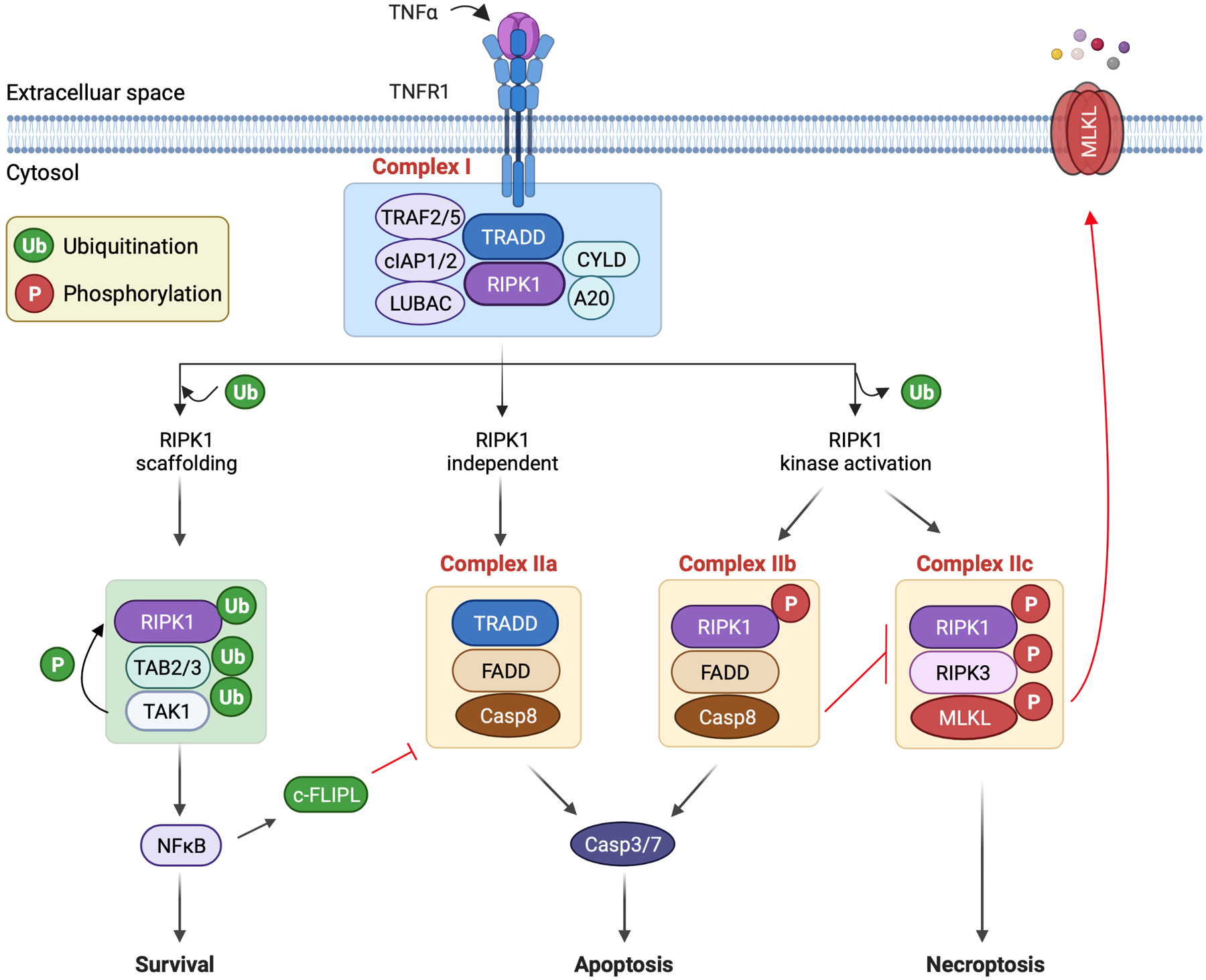
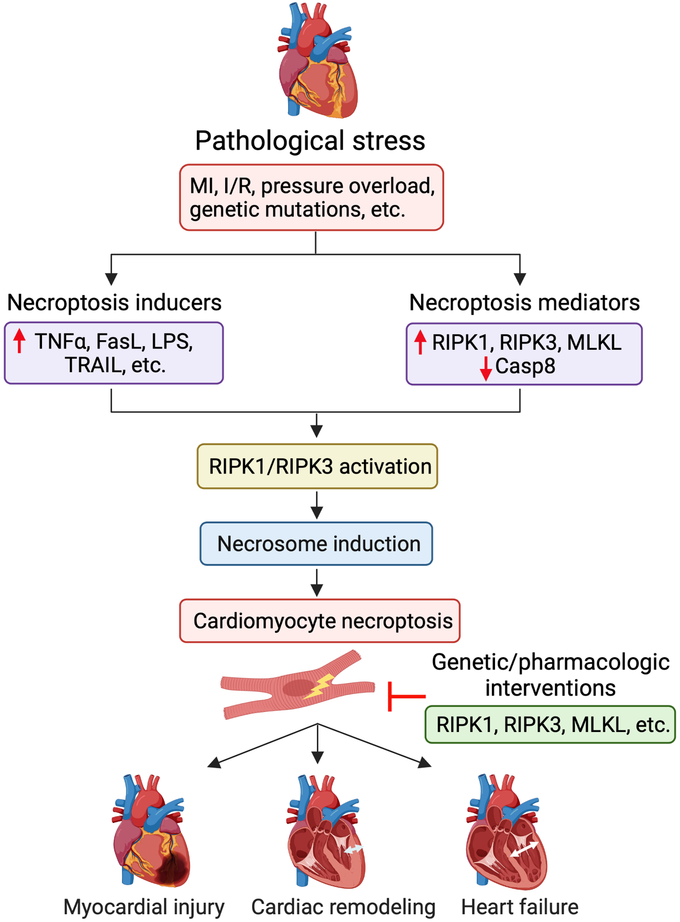
Cell death is a crucial event underlying cardiac ischemic injury, pathological remodeling, and heart failure. Unlike apoptosis, necrosis had long been regarded as a passive and unregulated process. However, recent studies demonstrate that a significant subset of necrotic cell death is actively mediated through regulated pathways - a process known as "regulated necrosis". As a form of regulated necrosis, necroptosis is mediated by death receptors and executed through the activation of receptor interacting protein kinase 3 (RIPK3) and its downstream substrate mixed lineage kinase-like domain (MLKL). Recent studies have provided compelling evidence that necroptosis plays an important role in myocardial homeostasis, ischemic injury, pathological remodeling, and heart failure. Moreover, it has been shown that genetic and pharmacological manipulations of the necroptosis signaling pathway elicit cardioprotective effects. Important progress has also been made regarding the molecular mechanisms that regulate necroptotic cell death in vitro and in vivo. In this review, we discuss molecular and cellular mechanisms of necroptosis, potential crosstalk between necroptosis and other cell death pathways, functional implications of necroptosis in heart disease, and new therapeutic strategies that target necroptosis signaling.

Keywords: Apoptosis; Heart failure; Myocardial infarction; Necroptosis; Necrosis.


Figures

Fig. 1.
Fig. 1.
Mechanisms of necroptosis. Necroptosis is initiated by the binding of death ligands to membrane-bound death receptors. Shown here is the binding of tumor necrosis factor-α (TNFα) to TNF receptor 1 (TNFR1). This initiates assembly of complex I on the cytoplasmic tail of the receptor. Complex I assembly may induce divergent cellular outcomes including cell survival, apoptosis, or necroptosis, which are mediated by distinct downstream cytosolic signaling complexes. In the formation of complex I, TNFR1 recruits the adaptor protein TNFR1-associated death domain protein (TRADD), which then recruits receptor interacting protein kinase 1 (RIPK1). TRADD also recruits the adaptor proteins TNF receptor-associated factor 2 and 5 (TRAF2/5) and cellular inhibitor of apoptosis proteins 1 and 2 (cIAP1/2), which catalyze lysine 63 (K63)-linked ubiquitination of RIPK1. The K63-linked ubiquitin chains also promote the recruitment of the linear ubiquitin chain assembly complex (LUBAC), which catalyzes linear [or methionine 1 (M1)-linked] ubiquitination of RIPK1. Also recruited into this complex are protein deubiquitinases cylindromatosis (CYLD) and A20, which mediate RIPK1 deubiquitination. The K63-linked ubiquitin chains on RIPK1 serve to recruit TAK1-binding protein 2 and 3 (TAB2 and TAB3) to facilitate the activation of transforming growth factor β-activated kinase-1 (TAK1). TAK1 promotes cell survival by activating nuclear factor-κB (NF-κB). TAK1 also prevents RIPK1 kinase activation through direct phosphorylation. Cell death initiation requires the transition from complex I to death-inducing cytosolic complexes. Complex IIa, which consists of TRADD-FADD-procaspase-8, signals RIPK1-independent apoptosis. In contrast, complex IIb, consisting of activated RIPK1-FADD-procaspase-8, mediates RIPK1-dependent apoptosis. Of note, caspase-8 inhibits necroptosis by cleaving RIPK1 and RIPK3. Moreover, in complex IIc (also termed “necrosome”), the binding of activated RIPK1 and RIPK3 leads to RIPK3 activation, which then phosphorylates and activates a pseudokinase called mixed lineage kinase-like domain (MLKL). MLKL undergoes oligomerization and translocates to and permeabilizes the plasma membrane to induce necroptosis.
Fig. 2.
Fig. 2.
Necroptosis in the pathogenesis of heart disease. Cardiac necroptosis signaling is activated in response to pathological stress (e.g., MI, I/R, pressure overload, and genetic mutations), a process that involves the induction of key necroptosis inducers (e.g., TNF, FasL, LPS, and TRAIL) and mediators (e.g., upregulation of RIPK1, RIPK3, and MLKL; downregulation of caspase-8). Activation of RIPK1 and/or RIPK3 is critical for the assembly of the necroptosis signaling complex (termed “necrosome”), which induces cell death by activating MLKL and subsequent plasma membrane rupture. Cardiomyocytes death via necroptosis plays an important role in the pathogenesis of myocardial ischemic injury, pathological remodeling, cardiac dysfunction, and heart failure. Genetic or pharmacologic interventions targeting necroptosis signaling proteins (e.g., RIPK1, RIPK3, and MLKL) represent potential therapeutic strategies for heart disease.

References

    1. Del Re DP, Amgalan D, Linkermann A, Liu Q, Kitsis RN, Fundamental mechanisms of regulated cell death and implications for heart disease, Physiol. Rev 99 (2019) 1765–1817. - PMC - PubMed
    1. Kung G, Konstantinidis K, Kitsis RN, Programmed necrosis, not apoptosis, in the heart, Circ. Res 108 (2011) 1017–1036. - PubMed
    1. Konstantinidis K, Whelan RS, Kitsis RN, Mechanisms of cell death in heart disease, Arterioscler. Thromb. Vasc. Biol 32 (2012) 1552–1562. - PMC - PubMed
    1. Whelan RS, Kaplinskiy V, Kitsis RN, Cell death in the pathogenesis of heart disease: mechanisms and significance, Annu. Rev. Physiol 72 (2010) 19–44. - PubMed
    1. Chen Z, Chua CC, Ho YS, Hamdy RC, Chua BH, Overexpression of Bcl-2 attenuates apoptosis and protects against myocardial I/R injury in transgenic mice, Am. J. Physiol. Heart Circ. Physiol 280 (2001) H2313–H2320. - PubMed

Publication types

Substances