

Optical biopsy for esophageal squamous cell neoplasia by using endocytoscopy
- PMID: 35597920
- PMCID: PMC9123668
- DOI: 10.1186/s12876-022-02335-5
Optical biopsy for esophageal squamous cell neoplasia by using endocytoscopy
Abstract
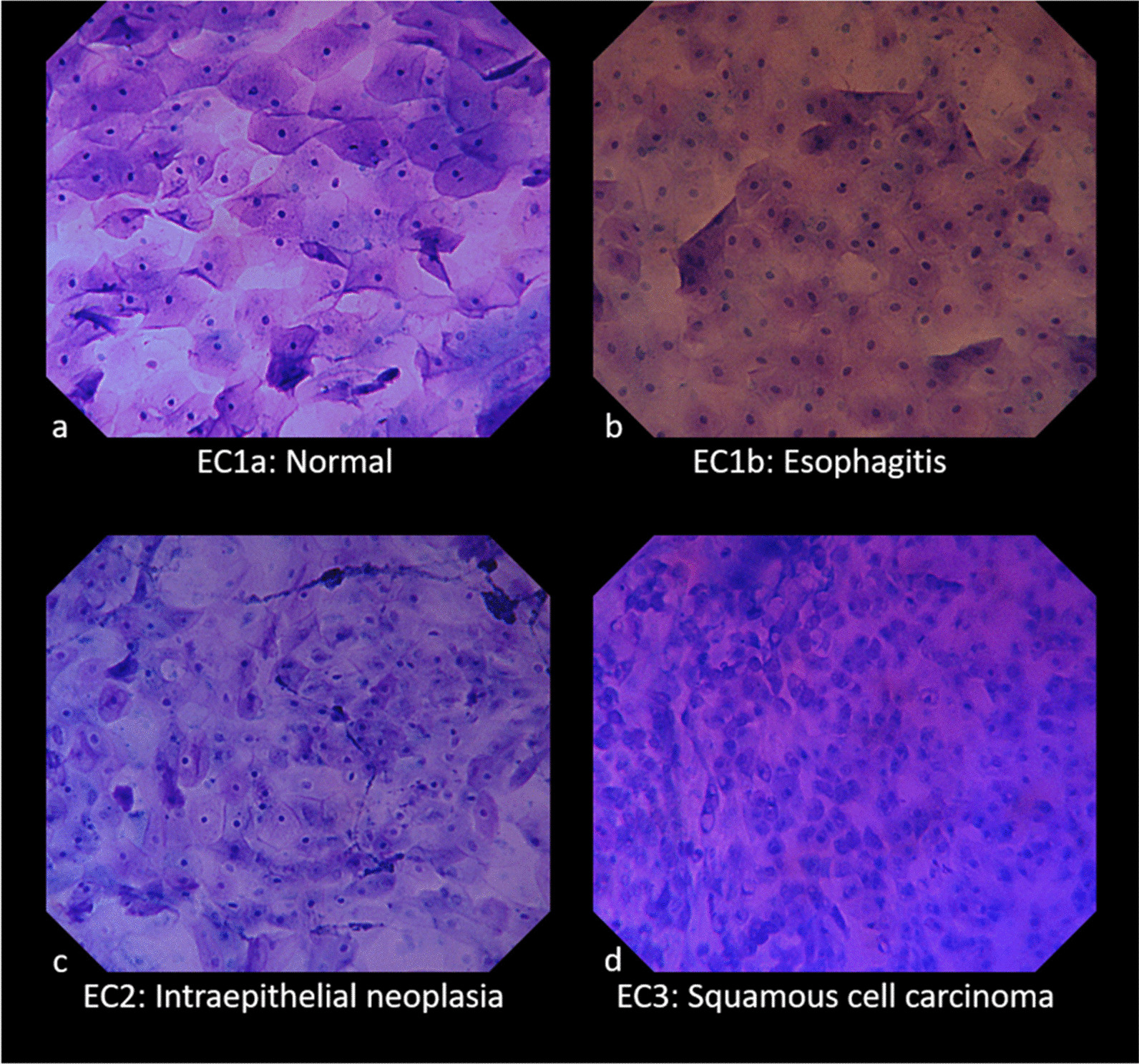
Background: Endocytoscopy (ECS) enables microscopic observation in vivo for the gastrointestinal mucosa; however, there has been no prospective study in which the diagnostic accuracy of ECS for lesions that have not yet undergone histological diagnosis was evaluated. We conducted a surveillance study for patients in a high-risk group of esophageal squamous cell carcinoma (ESCC) and evaluated the in vivo histological diagnostic accuracy of ECS.
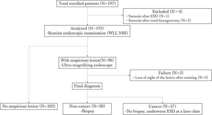
Methods: This study was a multicenter prospective study. We enrolled 197 patients in the study between September 1, 2019 and November 30, 2020. The patients first underwent white light imaging and narrow band imaging, and ultra-high magnifying observation was performed if there was a lesion suspected to be an esophageal tumor. Endoscopic submucosal dissection (ESD) was later performed for lesions that were diagnosed to be ESCC by ECS without biopsy. We evaluated the diagnostic accuracy of ECS for esophageal tumorous lesions.
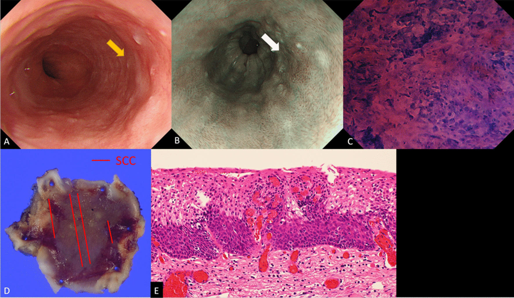
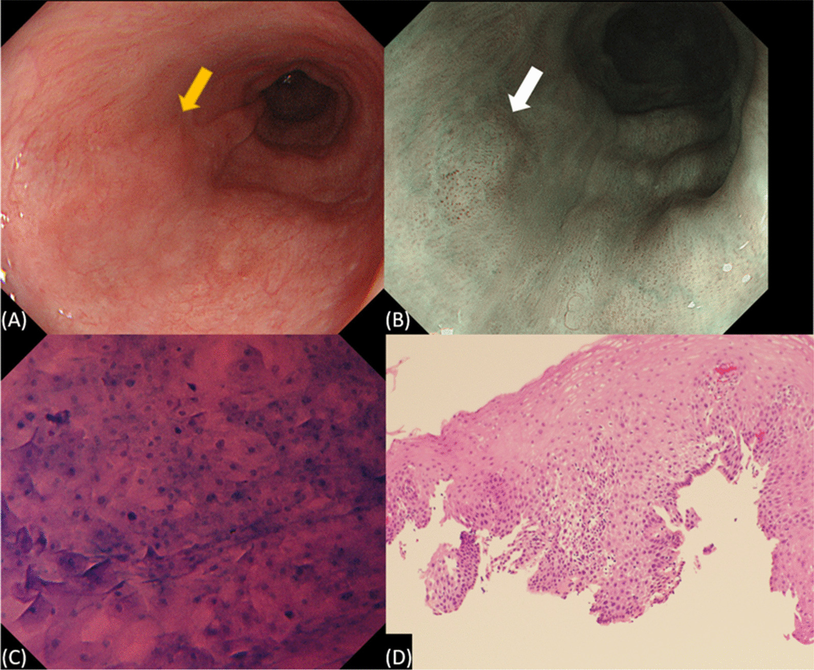
Results: ESD was performed for 37 patients (41 lesions) who were diagnosed as having ESCC by ECS, and all of them were histopathologically diagnosed as having ESCC. The sensitivity [95% confidence interval (CI)] was 97.6% (87.7-99.7%), specificity (95% CI) was 100% (92.7-100%), diagnostic accuracy (95% CI) was 98.9% (94.0-99.8%), positive predictive value (PPV) (95% CI) was 100% (91.4-100%) and negative predictive value (NPV) (95% CI) was 98.0% (89.5-99.7%).
Conclusions: ECS has a high diagnostic accuracy and there were no false positives in cases diagnosed and resected as ESCC. Optical biopsy by using ECS for esophageal lesions that are suspected to be tumorous is considered to be sufficient in clinical practice.
Keywords: Diagnostic accuracy; Endocytoscopy; Esophageal cancer; Intraepithelial neoplasia; Optical biopsy.
© 2022. The Author(s).
Conflict of interest statement
The authors have no conflict of interest to declare.
Figures
References
-
- Takahashi M, Shimizu Y, Ono M, Suzuki M, Omori S, Yoshida T, et al. Endoscopic diagnosis of early neoplasia of the esophagus with narrow band imaging: correlations among background coloration and iodine staining findings. J Gastroenterol Hepatol. 2014;29:762–768. doi: 10.1111/jgh.12477. - DOI - PubMed
-
- Nagami Y, Tominaga K, Machida H, Nakatani M, Kameda N, Sugimori S, et al. Usefulness of non-magnifying narrow-band imaging in screening of early esophageal squamous cell carcinoma: a prospective comparative study using propensity score matching. Am J Gastroenterol. 2014;109:845–854. doi: 10.1038/ajg.2014.94. - DOI - PMC - PubMed
Publication types
MeSH terms
LinkOut - more resources
Full Text Sources
Medical
Miscellaneous
