. 2022 May 21;53(1):36.
doi: 10.1186/s13567-022-01055-8.

Identification of excretory and secretory proteins from Haemonchus contortus inducing a Th9 immune response in goats

Affiliations

Identification of excretory and secretory proteins from Haemonchus contortus inducing a Th9 immune response in goats

Meng Liang et al. Vet Res. .

Abstract

Th9 cells have been shown to play crucial roles in anti-parasite immunity, pathogenic microbe infection, and allergy. Previous studies have demonstrated that Haemonchus contortus excretory and secretory proteins (HcESPs) induce the proliferation of Th9 cells and alter the transcriptional level of IL-9 as well as its related pathways in the Th9 immune response after infection. However, the exact molecule(s) in HcESPs inducing the Th9 immune response is not yet known. In this study, flow cytometry, co-immunoprecipitation (Co-IP) and shotgun liquid chromatography tandem-mass spectrometry (LC-MS/MS) were used, and a total of 218 proteins from HcESPs that might interact with goat Th9 cells were identified. By in vitro culture of Th9 cells with HcESPs, 40 binding proteins were identified. In vivo, 38, 47, 42 and 142 binding proteins were identified at 7, 15, 35 and 50 days post-infection (dpi), respectively. Furthermore, 2 of the 218 HcESPs, named DNA/RNA helicase domain containing protein (HcDR) and GATA transcription factor (HcGATA), were confirmed to induce the proliferation of Th9 cells and promote the expression of IL-9 when incubated with goat peripheral blood mononuclear cells (PBMCs). This study represents a proteomics-guided investigation of the interactions between Th9 cells and HcESPs. It provides a new way to explore immunostimulatory antigens among HcESPs and identifies candidates for immune-mediated prevention of H. contortus infection.

Keywords: Haemonchus contortus; HcDR; HcGATA; Th9 immune response; binding molecules; proteomics.


Conflict of interest statement

The authors declare that they have no competing interests.

Figures

Figure 1
Figure 1
Collection of HcESPs and preparation of anti-HcESPs rat IgG. Lane 1: SDS–PAGE showing HcESP production; Lanes 2, 3: Western blot showing HcESPs identified with anti-HcESPs rat IgG as the primary antibody and normal rat IgG as the negative control.
Figure 2
Figure 2
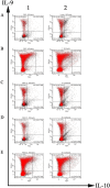
Th9-cell sorting by flow cytometry. A Th9 cells induced by HcESPs in vitro. PBMCs were treated with PBS (0 μg/mL HcESPs as the control) (A Panel 1) or 80 μg/mL HcESPs (A Panel 2). Th9 cells were sorted by flow cytometry using CD2 + CD4 + IL-9 + IL-10 + as the gate. BE Percentages of Th9 cells at 7 (B), 15 (C), 35 (D) and 50 (E) dpi. Samples were collected from the blank group (B–E Panel 1) and challenge group (B–E Panel 2) at 7, 15, 35 and 50 dpi.
Figure 3
Figure 3
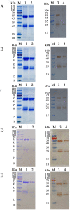
Co-IP assays indicated that HcESPs bind to Th9 cells. Lane M: marker. Lanes 1 and 2: SDS–PAGE showing that cell lysates were immunoprecipitated by anti-HcESPs rat IgG (Lane 1) or normal rat IgG as a negative control (Lane 2). Lanes 3 and 4: Western blot showing that cell lysates were immunoprecipitated by anti-HcESPs rat IgG (Lane 4) or normal rat IgG as a negative control (Lane 4). Cell lysates from in vitro experiments (A) or in vivo experiments collected at 7 (B), 15 (C), 35 (D) and 50 (E) dpi. Anti-ESPs rat IgG was used in the Western blot analysis to identify proteins that bound to Th9 cells.
Figure 4
Figure 4
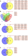
Venn diagram of binding proteins. A The binding proteins shared between in vitro and in vivo experiments. B The binding proteins shared among different developmental stages (7, 15, 35 and 50 dpi). C The binding proteins shared between 7 and 15 dpi. D The binding proteins shared between 15 and 35 dpi. E The binding proteins shared in the in vivo experiment between 35 and 50 dpi.
Figure 5
Figure 5
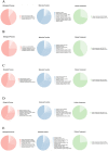
GO annotation. A HcESPs that bound to goat Th9 cells in vitro. B HcESPs identified at 7 dpi. C HcESPs identified at 15 dpi. D HcESPs identified at 35 dpi. E HcESPs identified at 50 dpi.
Figure 6
Figure 6
Effects of rHcDR on Th9 cells and IL-9 expression in vitro. A Agarose gel electrophoresis of the HcDR gene. Lane 1: amplification products of the HcDR gene. Lane M: DNA molecular weight marker. B Purification of rHcDR. Lane 2: before purification. Lane 3: purified rHcDR. Lane M: standard protein molecular marker. C Western blot. Lane 4: rHcDR recognized by serum from an H. contortus-infected goat. Lane 5: No recognition by normal serum. Lane M: standard protein molecular marker. D The effects of rHcDR on Th9-cell proliferation. Goat PBMCs were treated with different concentrations of rHcDR (0, 5, 10, 20, 40 and 60 μg/mL). Th9 cells were detected by flow cytometry using staining with antibodies specific for typical intracellular cytokines (IL-9 and IL-10). E Proportions of Th9 cells observed with different concentrations of rHcDR (0, 5, 10, 20, 40 and 60 μg/mL). Data are presented as the mean ± SD and are representative of triplicate experiments (*p < 0.05, ****p < 0.0001). F Fold change in relative IL-9 mRNA expression. Goat PBMCs were stimulated with different concentrations of rHcDR. The significance level was set at *p < 0.05, **p < 0.01, or ****p < 0.0001, and “ns” indicates non-significance compared with the control (blank). Data are representative of three independent experiments.
Figure 7
Figure 7
Effects of rHcGATA on Th9-cell proliferation and IL-9 transcription. A Agarose gel electrophoresis of the HcGATA gene. Lane 1: reverse transcription PCR products of HcGATA; Lane M: DNA molecular weight marker. B Purification of rHcGATA. Lane 2: rHcGATA before purification. Lane 3: purified rHcGATA. Lane M: standard protein molecular marker. C Western blot. Lane 4: rHcGATA protein recognized by serum from an H. contortus-infected goat. Lane 5: rHcGATA was not recognized by normal serum. Lane M: standard protein molecular marker. D The effects of HcGATA on the proliferation of Th9 cells in vitro. PBMC-derived Th9 cells treated with a control (0 μg/mL) or different concentrations of HcGATA (5, 10, 20, 40, 60 μg/mL) were tested by flow cytometry using antibodies specific for typical intracellular cytokines (IL-9 and IL-10). E Proportions of Th9 cells observed with different concentrations of HcGATA (0, 5, 10, 20, 40 and 60 μg/mL). Data are presented as the mean ± SD representative of triplicate experiments (ns p > 0.05, *p < 0.05, ****p < 0.0001). F Fold change in relative IL-9 mRNA expression. Goat PBMCs were stimulated with different concentrations of rHcGATA. The significance level was set at ***p < 0.001, or ****p < 0.0001, and “ns” indicates non-significance compared with the control group. Data are representative of three independent experiments.

Similar articles

Cited by

References

    1. Kaplan RM, Vidyashankar AN. An inconvenient truth: global worming and anthelmintic resistance. Vet Parasitol. 2012;186:70–78. - PubMed
    1. Floate KD. Endectocide use in cattle and fecal residues: environmental effects in Canada. Can J Vet Res. 2006;70:1–10. - PMC - PubMed
    1. Geurden T, Chartier C, Fanke J, di Regalbono AF, Traversa D, von Samson-Himmelstjerna G, Demeler J, Vanimisetti HB, Bartram DJ, Denwood MJ. Anthelmintic resistance to ivermectin and moxidectin in gastrointestinal nematodes of cattle in Europe. Int J Parasitol Drugs Drug Resist. 2015;5:163–1714. - PMC - PubMed
    1. Hu Y, Cheng H. Health risk from veterinary antimicrobial use in China’s food animal production and its reduction. Environ Pollut. 2016;219:993–997. - PubMed
    1. Nisbet AJ, Meeusen EN, González JF, Piedrafita DM. Immunity to Haemonchus contortus and vaccine development. Adv Parasitol. 2016;93:353–396. - PubMed

LinkOut - more resources