

Papillary muscle avulsion following balloon mitral valvotomy evaluated with three-dimensional echocardiography
- PMID: 35597976
- PMCID: PMC9123678
- DOI: 10.1186/s12893-022-01636-6
Papillary muscle avulsion following balloon mitral valvotomy evaluated with three-dimensional echocardiography
Abstract
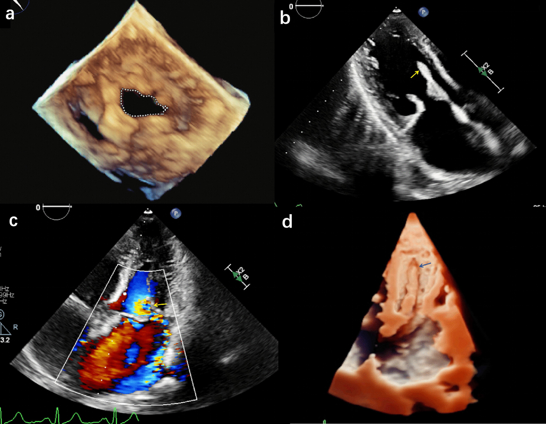
Background: Percutaneous balloon mitral valvotomy is a common therapeutic approach for rheumatic mitral stenosis. Avulsion of the papillary muscle is a rare but serious complication of balloon mitral valvotomy. The papillary muscles are derived from the trabecular layer of the developing ventricular walls. When subjected to a force, avulsion of papillary muscle from the trabecular layer may occur.
Case presentation: In this case report, we describe a patient with rheumatic mitral stenosis, who experienced avulsion of the mitral papillary muscle from the left ventricular wall after undergoing balloon mitral valvotomy. Papillary muscle alvusion resulted in severe mitral regurgitation, which was finally treated by mitral valve replacement.
Conclusion: We successfully diagnosed avulsion of the papillary muscle following balloon mitral valvotomy. Three-dimensional transthoracic echocardiography provides more information on mitral apparatus structure than two-dimensional transthoracic echocardiography.
Keywords: Alvusion; Balloon mitral valvotomy; Papillary muscle.
© 2022. The Author(s).
Conflict of interest statement
The authors declare that they have no competing interest.
Figures
References
Publication types
MeSH terms
LinkOut - more resources
Full Text Sources
