Review
. 2022 May 21;15(1):118.
doi: 10.1186/s12920-022-01267-z.

Multiple metastases of androgen indifferent prostate cancer in the urinary tract: two case reports and a literature review

Affiliations
Review

Multiple metastases of androgen indifferent prostate cancer in the urinary tract: two case reports and a literature review

Tsukasa Masuda et al. BMC Med Genomics. .

Abstract

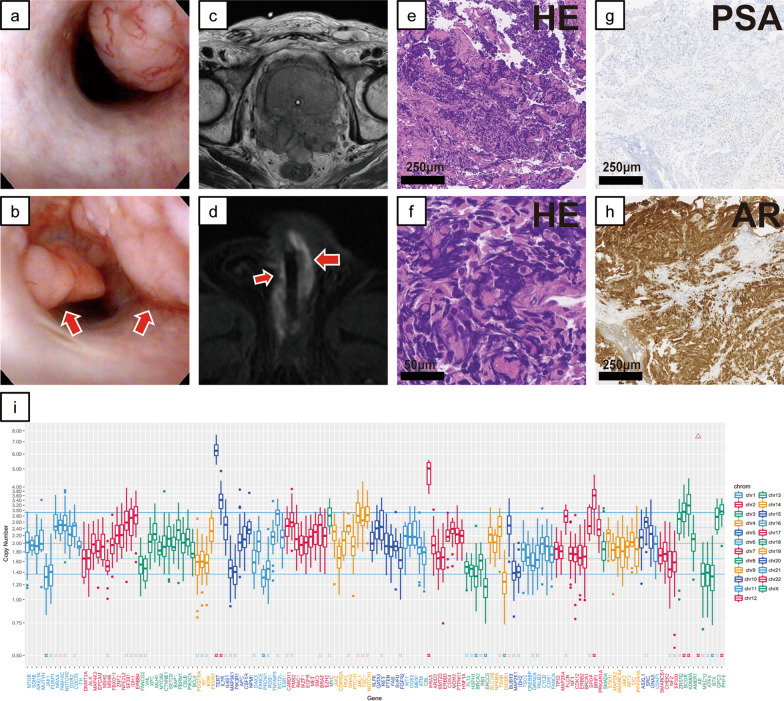
Background: Prostate cancer (PC) is mainly known to metastasize to bone, lung and liver, but isolated metastases of prostate cancer, including ductal carcinoma, in the urinary tract are very rare. We describe two patients with nodular masses in the urinary tract (the anterior urethra or the urinary bladder) that were found on cystoscopy during treatment of castration-resistant prostate cancer.

Case presentation: In both cases, the pathological diagnosis from transurethral tumor resection showed that they were androgen indifferent prostate cancer (AIPC), including aggressive variant prostate cancer (AVPC) in Case 1 and treatment-induced neuroendocrine differentiation prostate cancer (NEPC) in Case 2. In Case 1, Loss of genetic heterozygosity (LOH) of BRCA2 and gene amplification of KRAS was identified from the urethra polyps. In Case 2, homozygous deletion was observed in PTEN, and LOH without mutation was observed in RB1.

Conclusion: These are the first reports of two cases of urinary tract metastasis of AIPC.

Keywords: AR; Aggressive variant prostate cancer; BRCA2; Neuroendocrine differentiation prostate cancer; PTEN; TP53; Urinary tract metastasis.


Conflict of interest statement

The authors declare no competing interests.

Figures

Fig. 1
Fig. 1
Cystoscopic, imaging, and pathological examination results and genomic sequencing in Case 1. a, b Cystoscopic findings in the urethra. The cystoscope shows several nodular polyps in the proximal penile urethra and distal bulbar urethra. c Prostate magnetic resonance imaging (MRI). The prostate was almost entirely replaced by the tumor, which has invaded the rectum. d MR image of the urethra. Metastases of the prostate cancer extended with skip lesions along the corpus spongiosum in the entire anterior urethra. eh Representative microscopic images of hematoxylin and eosin (HE) staining and prostate-specific antigen and androgen receptor immunohistochemical staining of transurethral resections of urethra tumor specimens. These images were obtained using the following equipment: microscope, BX53; objective lens, UPLXAPO; camera, DP27; adapter, U-TV1XC. NanoZoomer-XR C12000 was used as acquisition software and the measured resolution was 500 dpi. i Examined genes (horizontal axis) and the copy number in Case 1 (vertical axis)
Fig. 2
Fig. 2
Cystoscopic, imaging, and pathological examination results and genomic sequencing in Case 2. a Bladder magnetic resonance imaging. b Pelvic computed tomography. Non-muscle-invasive masses in the neck and posterior wall of the urinary bladder are shown. c, d Cystoscopic findings. The tumor invaded from the prostate gland and has obstructed the bladder neck (c) and nodular masses are shown at the left posterior bladder wall (d). eh Representative microscopic images of hematoxylin and eosin (HE) staining and prostate-specific antigen, androgen receptor, and synaptophysin immunohistochemical staining and transurethral resection of the bladder tumor specimens. These images were obtained using the following equipment: microscope, BX53; objective lens, UPLXAPO; camera, DP27; adapter, U-TV1XC. NanoZoomer-XR C12000 was used as acquisition software and the measured resolution was 500 dpi. i Examined genes (horizontal axis) and the copy number in Case 2 (vertical axis)

References

    1. Conteduca V, et al. Clinical features of neuroendocrine prostate cancer. Eur J Cancer. 2019;121:7–18. doi: 10.1016/j.ejca.2019.08.011. - DOI - PMC - PubMed
    1. Berchuck JE, Viscuse PV, Beltran H, Aparicio A. Clinical considerations for the management of androgen indifferent prostate cancer. Prostate Cancer Prostatic Dis. 2021;24:623–637. doi: 10.1038/s41391-021-00332-5. - DOI - PMC - PubMed
    1. Aggarwal R, et al. Clinical and genomic characterization of treatment-emergent small-cell neuroendocrine prostate cancer: a multi-institutional prospective study. J Clin Oncol. 2018;36:2492–2503. doi: 10.1200/JCO.2017.77.6880. - DOI - PMC - PubMed
    1. Epstein JI, et al. Proposed morphologic classification of prostate cancer with neuroendocrine differentiation. Am J Surg Pathol. 2014;38:756–767. doi: 10.1097/PAS.0000000000000208. - DOI - PMC - PubMed
    1. Haller B, Yao HH-I, Christidis D, Chee J, Bishop C. Clinical recurrence of prostatic ductal adenocarcinoma in the anterior urethra. J Clin Urol. 2020 doi: 10.1177/2051415820921312. - DOI