

Slowly expanding lesions relate to persisting black-holes and clinical outcomes in relapse-onset multiple sclerosis
- PMID: 35598462
- PMCID: PMC9130104
- DOI: 10.1016/j.nicl.2022.103048
Slowly expanding lesions relate to persisting black-holes and clinical outcomes in relapse-onset multiple sclerosis
Abstract
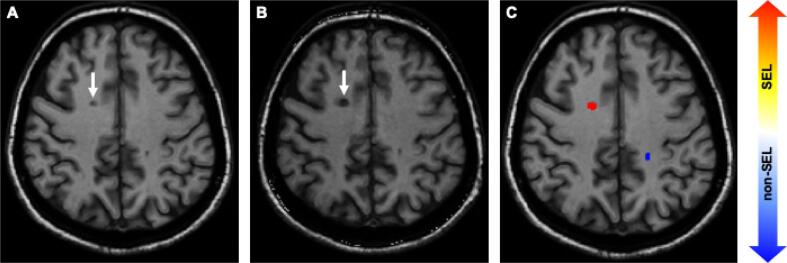
Background: Slowly expanding lesions (SELs) are MRI markers of chronic active lesions in multiple sclerosis (MS). T1-hypointense black holes, and reductions in magnetization transfer ratio (MTR) are pathologically correlated with myelin and axonal loss. While all associated with progressive MS, the relationship between these lesion's metrics and clinical outcomes in relapse-onset MS has not been widely investigated.
Objectives: To explore the relationship of SELs with T1-hypointense black holes, and longitudinal T1 intensity contrast ratio and MTR, their correlation to brain volume, and their contribution to MS disability in relapse-onset patients.
Methods: 135 patients with relapsing-remitting MS (RRMS) were studied with clinical assessments and brain MRI (T2/FLAIR and T1-weighted scans at 1.5/3 T) at baseline and two subsequent follow-ups; a subset of 83 patients also had MTR acquisitions. Early-onset patients were defined when the baseline disease duration was ≤ 5 years (n = 85). SELs were identified using deformation field maps from the manually segmented baseline T2 lesions and differentiated from the non-SELs. Persisting black holes (PBHs) were defined as a subset of T2 lesions with a signal below a patient-specific grey matter T1 intensity in a semi-quantitative manner. SELs, PBH counts, and brain volume were computed, and their associations were assessed through Spearman and Pearson correlation. Clusters of patients according to low (up to 2), intermediate (3 to 10), or high (more than 10) SEL counts were determined with a Gaussian generalised mixture model. Mixed-effects and logistic regression models assessed volumes, T1 and MTR within SELs, and their correlation with Expanded Disability Status Scale (EDSS) and confirmed disability progression (CDP).
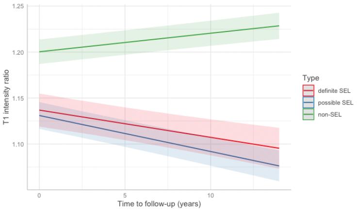
Results: Mean age at study onset was 35.5 years (73% female), disease duration 5.5 years and mean time to last follow-up 6.5 years (range 1 to 12.5); median baseline EDSS 1.5 (range 0 to 5.5) and a mean EDSS change of 0.31 units at final follow-up. Among 4007 T2 lesions, 27% were classified as SELs and 10% as PBHs. Most patients (n = 65) belonged to the cluster with an intermediate SEL count (3 to 10 SELs). The percentage of PBHs was higher in SELs than non-SELs (up to 61% vs 44%, p < 0.001) and within-patient SEL volumes positively correlated with PBH volumes (r = 0.53, p < 0.001). SELs showed a decrease in T1 intensity over time (beta = -0.004, 95%CI -0.005 to -0.003, p < 0.001), accompanied by lower cross-sectional baseline and follow-up MTR. In mixed-effects models, EDSS worsening was predicted by the SEL log-volumes increase over time (beta = 0.11, 95%CI 0.03 to 0.20, p = 0.01), which was confirmed in the sub-cohort of patients with early onset MS (beta = 0.14, 95%CI 0.04 to 0.25, p = 0.008). In logistic regressions, a higher risk for CDP was associated with SEL volumes (OR = 5.15, 95%CI 1.60 to 16.60, p = 0.006).
Conclusions: SELs are associated with accumulation of more destructive pathology as indicated by an association with PBH volume, longitudinal reduction in T1 intensity and MTR. Higher SEL volumes are associated with clinical progression, while lower ones are associated with stability in relapse-onset MS.
Keywords: Black holes; Chronic active lesions; Multiple sclerosis; SEL; Volumetric MRI.
Copyright © 2022. Published by Elsevier Inc.
Conflict of interest statement
The authors declare that they have no known competing financial interests or personal relationships that could have appeared to influence the work reported in this paper.
Figures
References
-
- Prineas J.W., Kwon E.E., Cho E.-S., Sharer L.R., Barnett M.H., Oleszak E.L., Hoffman B., Morgan B.P. Immunopathology of secondary-progressive multiple sclerosis. Ann Neurol. 2001;50(5):646–657. - PubMed
-
- Barkhof F., Brück W., De Groot C.J.A., Bergers E., Hulshof S., Geurts J., Polman C.H., van der Valk P. Remyelinated Lesions in Multiple Sclerosis. Arch Neurol. 2003;60(8):1073. - PubMed
Publication types
MeSH terms
Grants and funding
LinkOut - more resources
Full Text Sources
Medical
