. 2022 Jul 1:494:140-151.
doi: 10.1016/j.neuroscience.2022.05.018. Epub 2022 May 20.

Layers 3 and 4 Neurons of the Bilateral Whisker-Barrel Cortex

Affiliations

Layers 3 and 4 Neurons of the Bilateral Whisker-Barrel Cortex

Vassiliy Tsytsarev et al. Neuroscience. .

Abstract

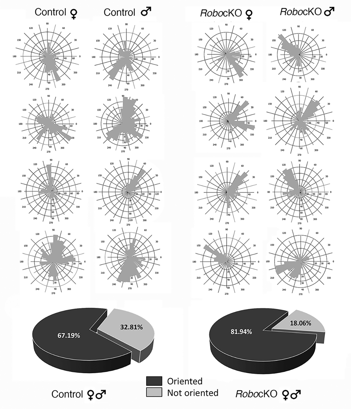
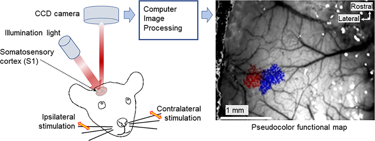
In Robo3R3-5cKO mouse brain, rhombomere 3-derived trigeminal principal nucleus (PrV) neurons project bilaterally to the somatosensory thalamus. As a consequence, whisker-specific neural modules (barreloids and barrels) representing whiskers on both sides of the face develop in the sensory thalamus and the primary somatosensory cortex. We examined the morphological complexity of layer 4 barrel cells, their postsynaptic partners in layer 3, and functional specificity of layer 3 pyramidal cells. Layer 4 spiny stellate cells form much smaller barrels and their dendritic fields are more focalized and less complex compared to controls, while layer 3 pyramidal cells did not show notable differences. Using in vivo 2-photon imaging of a genetically encoded fluorescent [Ca2+] sensor, we visualized neural activity in the normal and Robo3R3-5cKO barrel cortex in response to ipsi- and contralateral single whisker stimulation. Layer 3 neurons in control animals responded only to their contralateral whiskers, while in the mutant cortex layer 3 pyramidal neurons showed both ipsi- and contralateral whisker responses. These results indicate that bilateral whisker map inputs stimulate different but neighboring groups of layer 3 neurons which normally relay contralateral whisker-specific information to other cortical areas.

Keywords: Ca(2+)-fluorescence protein; In vivo 2-photon imaging; conditional Robo3 knockout; dendritic complexity; somatosensory cortex; whiskers.


Figures

Figure 1.
Figure 1.
Barrel cortex in control (A, C, E) and Robor3–5 cKO mice (B, D, F). A and B immunostained for VGlut2 (marker for TCAs, green) and NeuN (neurons, red). Insets show only NeuN immunostaining. Whisker barrel rows (a-e) are marked, in B asterisks outline the ipsilateral whisker representation area. In the same micrograph ipsilateral and contralateral c2 whisker barrels are indicated. C,D. Cytochrome oxidase (CO) staining reveals the barrel patterns in control (C) and knockout (D) mice. E, F. DAPI staining shows cellular barrel patterns. Scale bar in E = 500 μm.
Figure 2.
Figure 2.
Dendritic orientation plots of representative cells from female and male control and Robo3cKO mice. Pie charts show the percentages of cells showing orientation in control and mutant animals. (Control, 67.19%; Mutant, 81.94%, black) versus cells that are not oriented (Control, 32.81%; Mutant, 18.06%, gray).
Figure 3.
Figure 3.
Layer 4 spiny stellate neuron dendritic morphology analysis. Panel A shows drawings of layer 4 spiny stellate neurons, left to right 2 female, 2 male cells and top row from control cases, bottom row Robo3R3–5cKO cases. B and C show images of 4 spiny stellate neurons, with B from a control mouse and C from a Robo3R3–5cKO mouse. Scale bar in B and C: 10μm. D represents the number of intersections per distance from the soma (Sholl) in μm; ns for 10, 150–170μm; **p<0.01 for 20, 140μm; ***p<0.001 for 30–130μm. E represents the number of nodes per distance from the soma in μm; p values are on the graph, *p<0.05. Dendritic length per distance from soma is shown in F; ns for 10, 150,160μm; *p<0.05 for 140μm; ***p<0.001 for 20–130μm. Whisker box plots show total dendritic length in G ( ***p<0.001), total terminals in H (***p<0.001), soma size in I (*p<0.05) and number of branches per branch order in J (order 1 ns p>0.05; order 2 ***p<0.001; order 3 ***p<0.001; order 4 3 ***p<0.001). In D-F, controls are shown in black and Robo3R3–5cKO mice are shown in light grey. In G-J, controls are shown in black and Robo3R3–5cKO mice are shown in white.
Figure 4.
Figure 4.
Layer 3 pyramidal neuron dendritic morphology analysis. Panel A shows drawings of pyramidal neurons, with 2 cells from control and 2 cells from Robo3R3–5cKO mice. B. Micrographs of Golgi stained cells. Scale bar: 25 μm. C, Number of intersections per distance from the soma (Sholl) in μm, ns for 10–60μm, 110–170μm, *p<0.05 for 70, 90, 100, 180, 190, 230–260μm; ***p<0.001 for 80 and 220μm. D represents dendritic length per distance from the soma, ns for 30–60μm, 130–210μm, *p<0.05 for 10, 20, 70, 100–120, 220–300μm; **p<0.01 for 80 and 90μm. E-H. Whisker box plots show total dendritic length, total terminals, soma size, and the number of branches per branch order. There were no significant differences for E, F and G. In H *p<0.05 for order 1, **p<0.01 for order 5.
Figure 5.
Figure 5.
Left: intrinsic optical imaging experimental setup. Right: IOS data from a mutant animal; region of contralateral whisker responses shown in blue, ipsilateral in red.
Figure 6.
Figure 6.
A. Maps of whisker-evoked fluorescence change in L2/3 neurons from representative control mice, obtained by 2-photon calcium imaging of S1 cortex barrel area during ipsi- or contralateral C2 whisker stimulation. Each dot depicts one neuron. The magnitude of evoked ΔF/Fo is indicated by the color bar. Scale bar: 100 μm. B. Same as panel A but for Robo3R3–5 cKO mice. C. Histograms showing distribution of laterality index values for neurons that show a statistically significant response to either whisker, combined across control animals. Light color bars indicate data from all responsive neurons, and dark color bars data from neurons with statistically significant laterality index. D. Same as panel C but for Robo3R3–5 cKO mice.

Similar articles

Cited by

References

    1. Chédotal A (2014) Development and plasticity of commissural circuits: from locomotion to brain repair. Trends Neurosci 37(10):551–62. - PubMed
    1. Da Silva RV, Johannssen H, Wyss ,MT, Roome RB, Bourojeni FB, Stifani N, Marsh APL, Ryan MM, et al. (2018) DCC is required for the development of nociceptive topognosis in Mice and Humans. Cell Rep 22(5): 1105–14. - PubMed
    1. Erzurumlu RS, Gaspar P (2012) Development and critical period plasticity of the barrel cortex. Eur J Neurosci 35(10):1540–53. - PMC - PubMed
    1. Erzurumlu RS, Gaspar P (2020) how the barrel cortex became a working model for developmental plasticity: a historical perspective. J Neurosci 40(34):6460–6473. - PMC - PubMed
    1. Erzurumlu RS, Murakami Y, Rijli FM (2010) Mapping the face in the somatosensory brainstem. Nat Rev Neurosci 11(4):252–63. - PMC - PubMed

Publication types