. 2022 Jul 15:447:116071.
doi: 10.1016/j.taap.2022.116071. Epub 2022 May 20.

Biological effects of crude oil vapor. IV. Cardiovascular effects

Affiliations

Biological effects of crude oil vapor. IV. Cardiovascular effects

Kristine Krajnak et al. Toxicol Appl Pharmacol. .

Abstract

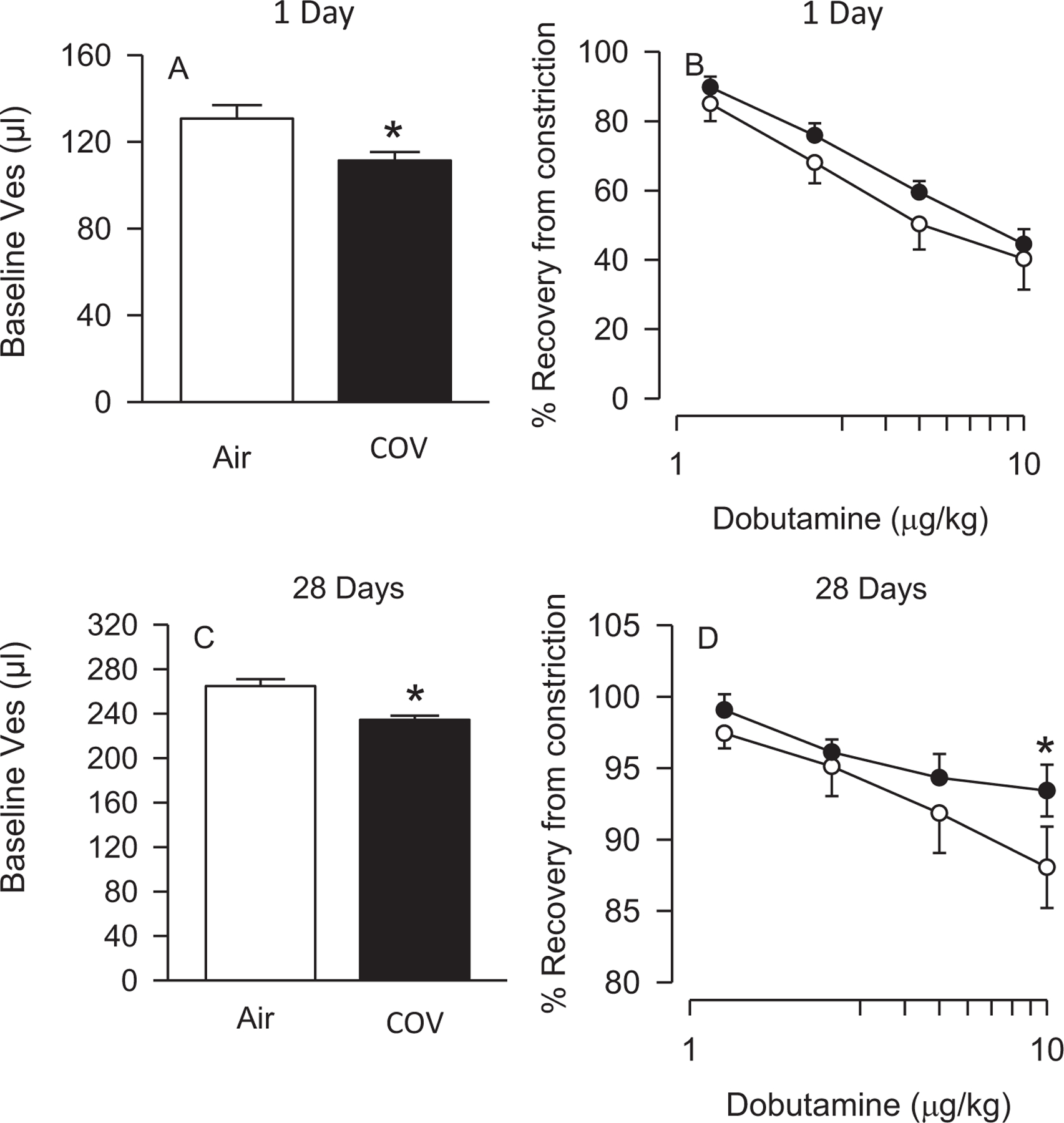
Workers in the oil and gas extraction industry are at risk of inhaling volatile organic compounds. Epidemiological studies suggest oil vapor inhalation may affect cardiovascular health. Thus, in this hazard identification study we investigated the effects of inhalation of crude oil vapor (COV) on cardiovascular function. Male rats were exposed to air or COV (300 ppm) for 6 h (acute), or 6 h/day × 4 d/wk. × 4 wk. (sub-chronic). The effects of COV inhalation were assessed 1, 28, and 90 d post-exposure. Acute exposure to COV resulted in reductions in mean arterial and diastolic blood pressures 1 and 28 d after exposure, changes in nitrate-nitrite and H2O2 levels, and in the expression of transcripts and proteins that regulate inflammation, vascular remodeling, and the synthesis of nitric oxide (NO) in the heart and kidneys. The sub-chronic exposure resulted in a reduced sensitivity to α1-adrenoreceptor-mediated vasoconstriction in vitro 28 d post-exposure, and a reduction in oxidative stress in the heart. Sub-chronic COV exposure led to alterations in the expression of NO synthases and anti-oxidant enzymes, which regulate inflammation and oxidative stress in the heart and kidneys. There seems to be a balance between changes in the expression of transcripts associated with the generation of reactive oxygen species (ROS) and antioxidant enzymes. The ability of antioxidant enzymes to reduce or inhibit the effects of ROS may allow the cardiovascular system to adapt to acute COV exposures. However, sub-chronic exposures may result in longer-lasting negative health consequences on the cardiovascular system.

Keywords: Adrenoreceptor modulation; Cardiovascular; Inflammation; Oxidative stress; Peripheral vascular; Renal.


Conflict of interest statement

Declaration of Competing Interest

The authors declare that they have no conflicts of interest in relation to this publication.

Figures

Fig. 1.
Fig. 1.
Fig. A and B show the dose-dependent vasoconstriction in the tail artery of rats in response to phenylephrine (PE) 1 d (A) and 28 d (B), and acetylcholine (ACh) 1 d (C) and 28 d (D) after an acute exposure to air or COV (n = 8 animals/group). No significant changes in vascular responses to either PE-induced vasoconstriction or ACh-induced re-dilation in response to treatment or days of recovery were observed.
Fig. 2.
Fig. 2.
Fig 2. A-C show the dose-dependent vasoconstriction in response to PE-induced vasoconstriction 1 d (A), 28 d (B) and 90 d (C) after a sub-chronic exposure to COV in the ventral tail arteries of rats. n = 5–8 animals/group. Twenty-eight days following exposure, arteries from COV-exposed rats were less sensitive to PE-induced vasoconstriction than those from air-exposed animals (*p < 0.05). Fig. D-F show responses to ACh-induced re-dilation in the same arteries. There were no effects of air or COV on ACh-induced re-dilation.
Fig. 3.
Fig. 3.
The effects of COV inhalation on ventricular systolic-end pressure (Ves) and diastolic-end pressure (Ved). One d following an acute exposure, baseline Ves was reduced, and 28 d following exposure Ved was reduced in COV-exposed rats (A and C, respectively). Dobutamine-induced recovery from vasoconstriction was not affected 1 d following COV exposure (Fig. 3B). However, 28 d following COV exposure, dobutamine-induced recovery from vasoconstriction was reduced as compared to controls (Fig. 3D).
Fig. 4.
Fig. 4.
Diastolic blood pressure (DBP) and mean arterial blood pressure (MAP) measured by the PV-loop method after an acute exposure to air or COV. One d following exposure, both DBP (A) and MAP (B) were significantly reduced as compared to air controls. *p < 0.05. n = 8 animals/group.
Fig. 5.
Fig. 5.
Nox concentrations in the heart (A, B) and kidney (C, D) 1 and 28 d after an acute exposure to air or COV. In the heart, Nox concentrations were significantly increased 28 d after exposure to COV (A). However, there were not significant differences in Nox expression in the kidney (B) after exposure to COV. However, H2O2 concentrations were significantly increased 1 d after COV exposure in the heart and reduced 27 d following exposure in the kidney. *p < 0.05, as compared to time-matched controls. n = 8 animals/group.
Fig. 6.
Fig. 6.
Reactive oxygen species (ROS) concentrations in the heart (A) and kidney (B) of rats 1, 28 or 90 d following a sub-chronic exposure to air or COV. In the heart, ROS concentrations in air and COV exposed animals were not significantly different 1 and 90 d after exposure to COV. However, 28 d after exposure, air control ROS levels were less than levels on 1 d (* p < 0.05). ROS concentration in the 28 d after COV exposure were lower than air controls (# p < 0.05) and lower than ROS concentrations in COV animals after 1 and 90 d of exposure. In the kidney (B), ROS concentrations were variable, but there were no significant COV exposure-related changes in ROS on any day of the study. n = 8 animals/group.
Fig. 7.
Fig. 7.
Correlations between changes in different proteins in a protein panel for factors implicated in the development of cardiovascular disease after acute exposure to COV using multivariate regression analysis. Protein pairs are listed on the left, and bars with asterisks indicate that there was a significant correlation between changes in those proteins (p < 0.05). If there is a bar in the graphs, it indicates that there were significant correlations in the changes in that pair of proteins in both the air- and COV-treated animals. Correlations proteins measured in tissue collected 1 d (A) and 28 d (B) following acute COV exposure, and 1d (C) and 28 d (D) following sub-chronic COV exposure are presented. n = 6–8 animals/group.
Fig. 8.
Fig. 8.
Correlations between changes in different proteins on a protein panel, measuring changes in proteins indicative of kidney injury after acute exposure to COV. Protein pairs are listed on the left, and bars with an asterisk indicate that there was a significant correlation between changes in those proteins (p < 0.05). If there is an asterisk over a bar in the graph, it indicates that there were significant correlations in the protein changes in both the air- and COV-treated animals. Correlations between proteins measured in tissue collected 1d (A) and 28 d (B) following acute COV exposure, and 1d (C) and 28 d (D) following sub-chronic COV exposure are presented. n = 6–8 animals/group.

References

    1. Aguilera F, Mendez J, Pasaro E, Laffon B, 2010. Review on the effects of exposure to spilled oils on human health. J. Appl. Toxicol 30 (4), 291–301. - PubMed
    1. American Petroleum Institute, 2008. The Relationship between the Aromatic Ring Class Content and Selected Endpoints of Repeat-Dose and Developmental Toxicity of High-Boiling Petroleum Substances. cited Available from. http://www.petroleumhpv.org/~/media/PetroleumHPV/Documents/Publications/....
    1. Anttila A, Pokhrel A, Heikkila P, Viinanen R, Pukkala E, 2015. Kidney cancer risk in oil refining in Finland: a nested case-referent study. J. Occup. Environ. Med 57 (1), 68–72. - PubMed
    1. Aragon M, Erdely A, Bishop L, Salmen R, Weaver J, Liu J, Hall P, Eye T, Kodali V, Zeidler-Erdely P, Stafflinger JE, Ottens AK, Campen MJ, 2016. MMP-9-dependent serum-borne bioactivity caused by multiwalled carbon nanotube exposure induces vascular dysfunction via the CD36 scavenger receptor. Toxicol. Sci 150 (2), 488–498. - PMC - PubMed
    1. Bicu M, Mota M, Panduru NM, Graunteanu C, Mota E, 2010. Oxidative stress in diabetic kidney disease. Rom. J. Intern. Med 48 (4), 307–312. - PubMed

Publication types