. 2022 Aug:62:101516.
doi: 10.1016/j.molmet.2022.101516. Epub 2022 May 20.

Multiple metabolic pathways fuel the truncated tricarboxylic acid cycle of the prostate to sustain constant citrate production and secretion

Affiliations

Multiple metabolic pathways fuel the truncated tricarboxylic acid cycle of the prostate to sustain constant citrate production and secretion

Lilianne Frégeau-Proulx et al. Mol Metab. 2022 Aug.

Abstract

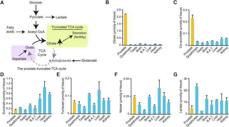
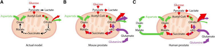
Objective: The prostate is metabolically unique: it produces high levels of citrate for secretion via a truncated tricarboxylic acid (TCA) cycle to maintain male fertility. In prostate cancer (PCa), this phenotype is reprogrammed, making it an interesting therapeutic target. However, how the truncated prostate TCA cycle works is still not completely understood.

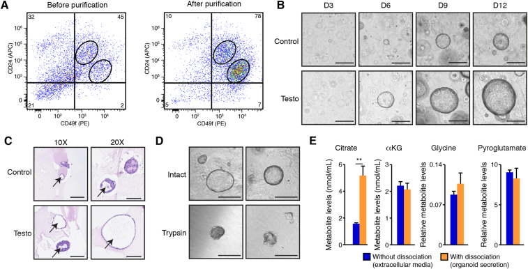
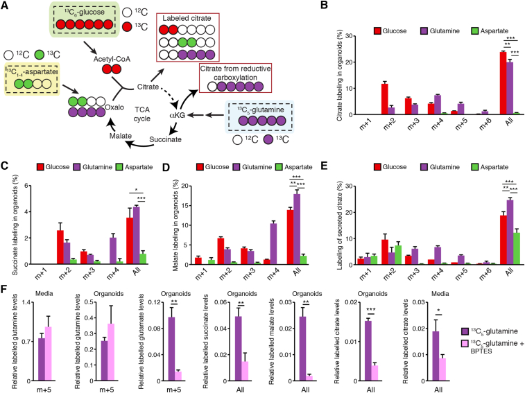
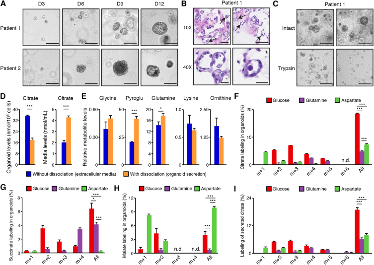
Methods: We optimized targeted metabolomics in mouse and human organoid models in ex vivo primary culture. We then used stable isotope tracer analyses to identify the pathways that fuel citrate synthesis.

Results: First, mouse and human organoids were shown to recapitulate the unique citrate-secretory program of the prostate, thus representing a novel model that reproduces this unusual metabolic profile. Using stable isotope tracer analysis, several key nutrients were shown to allow the completion of the prostate TCA cycle, revealing a much more complex metabolic profile than originally anticipated. Indeed, along with the known pathway of aspartate replenishing oxaloacetate, glutamine was shown to fuel citrate synthesis through both glutaminolysis and reductive carboxylation in a GLS1-dependent manner. In human organoids, aspartate entered the TCA cycle at the malate entry point, upstream of oxaloacetate. Our results demonstrate that the citrate-secretory phenotype of prostate organoids is supported by the known aspartate-oxaloacetate-citrate pathway, but also by at least three additional pathways: glutaminolysis, reductive carboxylation, and aspartate-malate conversion.

Conclusions: Our results add a significant new dimension to the prostate citrate-secretory phenotype, with at least four distinct pathways being involved in citrate synthesis. Better understanding this distinctive citrate metabolic program will have applications in both male fertility as well as in the development of novel targeted anti-metabolic therapies for PCa.

Keywords: Androgen; Fertility; Organoids; Prostate cancer; TCA cycle.


Figures

Figure 1
Figure 1
The prostate exhibits a unique truncated TCA cycle that supports massive citrate production. A) Current working model of the prostate's truncated TCA cycle. Citrate is massively produced by prostate epithelial cells and secreted into the seminal fluid; this is a well-known phenotype associated with fertility. Several nutrients could fuel the prostate TCA cycle, including glucose, fatty acids, glutamate, and aspartate. It is currently assumed that glucose and aspartate are the major carbon sources required for citrate production and secretion. (B–G) Targeted metabolomics of TCA cycle intermediates and lactate in various mouse tissues. Results are shown as the average ± SEM of two independent experiments comprising organs from seven mice. Results are shown for the prostate (yellow) and other organs (blue) in μmol/g of tissues. BAT: Brown adipose tissue. Additional metabolites quantified are available in Supplemental Figure S1.
Figure 2
Figure 2
Primary mouse prostate organoids exhibit the citrate-secretory phenotype. A) Flow cytometry analyses of basal epithelial cells (CD49fhigh/CD24intermediate) and luminal epithelial cells (CD49fintermediate/CD24high) before and after purification. Numbers in the corners are percentages. One representative experiment of two independent experiments is shown. B) Brightfield visualization of organoids over 12 days in three-dimensional culture with and without testosterone, an androgen treatment. Testosterone induces a significant increase in organoid size without changing the number of organoids (quantification of organoid numbers and sizes is available in Supplemental Figure S2C). Testo: Testosterone treatment. Scale bars = 300 μm. C) H&E visualization of mouse prostate organoids after 14 days in culture, with and without testosterone. Arrows show the presence of internal lumen in these organoids. Scale bars = 125 μm and 100 μm, respectively, for 10× and 20× view. Note that during the fixation process, bigger organoids tend to lose their circular architecture. D) Brief treatment with trypsin (3 min) disrupts the three-dimensional architecture of mouse prostate organoids to allow connection of the internal lumen with the extracellular media. Scale bars = 300 μm. E) GC–MS metabolite quantification in extracellular culture media of mouse prostate organoids treated with testosterone, with and without dissociation, as shown in D. With dissociation, this media contains luminal secretion. Results are shown as the average ± SEM of one representative experiment performed in quadruplicates, out of five independent experiments. ∗∗p < 0.01.
Figure 3
Figure 3
Contribution of glucose, glutamine, and aspartate to the prostate organoid TCA cycle. A) Schematic representation of respective carbon contributions when organoids are supplemented with 13C6-glucose, 13C5-glutamine, or 13C1-4-aspartate, assuming aspartate carbons enter the cycle via oxaloacetate. For graphical purposes, aspartate is shown as 13C2-aspartate, but labeling in B–E can be observed from 13C1-, 13C2-, 13C3-, or 13C4-aspartate. Stable isotope tracer analyses in organoids using in parallel these three tracers are then shown for citrate (B), succinate (C), and malate (D), as well as for extracellular levels of citrate (E). After 11 days in culture, organoids were incubated for 72 h with 13C6-glucose, 13C5-glutamine, or 13C1-4-aspartate before being dissociated and harvested for GC–MS analyses. “All” indicates the sum of all shown isotopomers for a given metabolite. Statistics were only calculated using the sum of all isotopomers of a given metabolite. Results are shown as the average ± SEM of one representative experiment performed in triplicate out of three independent experiments. For these tracer experiments, note that organoids were cultured in parallel with the different 13C-labeled nutrients. Secreted succinate and malate labeling is shown in Supplemental Figure S3. F) Stable isotope tracer analysis in organoids following 72 h exposure to 13C5-glutamine, with and without the GLS1 inhibitor BPTES. After 72 h, organoids and luminal secretion (media) were harvested for metabolomics. For glutamine and glutamate, m + 5 labeling is shown; for TCA cycle intermediates, as labeling patterns were more complex (as shown in B–E), the sum of total labeling is shown (as shown in B–E). Results are shown as the average ± SEM of one representative experiment performed in triplicates out of two independent experiments. ∗∗∗p < 0.001; ∗∗p < 0.01; ∗p < 0.05.
Figure 4
Figure 4
Metabolomics analyses in human prostate organoids. A) Brightfield visualization of normal human prostate organoids over 12 days in three-dimensional culture. Organoid cultures derived from two different human patients are shown. Scale bars = 200 μm. B) H&E visualization of patient-derived prostate organoids showing the presence of internal lumen, demonstrated by black arrows. Scale bars = 125 μm and 50 μm, respectively, for 10× and 40× view. C) Brief treatment with trypsin (3 min) disrupts the three-dimensional architecture of human prostate organoids to allow connection of the internal lumen with the extracellular media. Scale bars = 200 μm. Representative images from patient #1 are shown and images for patient-derived organoids from patient #2 are available in Supplemental Figure S4C. D) Intra-organoid and extracellular citrate level quantification by GC–MS, with and without dissociation as shown in C. With dissociation, media contains luminal secretion. E) GC–MS metabolite quantification in extracellular culture media of human prostate organoids, with and without dissociation, as shown in C. For D and E, results are shown as the average ± SEM of one representative experiment performed in triplicate out of three independent experiments. Pyroglu: pyroglutamate. Stable isotope tracer analysis in organoids using 13C6-glucose, 13C5-glutamine, or 13C1-4-aspartate tracers is then shown for intra-organoid citrate (F), succinate (G), and malate (H) levels, as well as for secreted citrate levels (I). For results shown in F–I, organoids were dissociated before GC–MS metabolomics quantifications. Results are shown as the average ± SEM of one representative experiment performed in triplicates out of two independent experiments for patient #1. For these tracer experiments, note that organoids were cultured in parallel with the different 13C-labeled nutrients. Similar results were obtained with organoids from patient #2 and are shown in Supplemental Figure S4. N.d.: not detectable or below the limit of quantification. “All” indicates the sum of all shown isotopomers for a given metabolite. Statistics were only calculated using the sum of all isotopomers of a given metabolite. ∗∗∗p < 0.001; ∗∗p < 0.01; ∗p < 0.05.
Figure 5
Figure 5
Updated working model of citrate metabolism in mouse and human prostate. A) The current working model of prostate metabolism describes a truncated TCA cycle that allows massive citrate secretion into the semen. In this model, glucose and aspartate are both essential for acetyl-CoA and oxaloacetate synthesis to sustain citrate synthesis by citrate synthase. However, metabolomics using mouse prostate tissues and mouse and human prostate organoids showed herein dress a more complex profile in the mouse (B) and human (C) prostate. First, aspartate is not essential for citrate synthesis. Second, citrate, but also downstream intermediates, can be produced using glucose, aspartate, and glutamine as carbon sources. Glutamine was revealed to be a significant nutrient contributing to citrate secretion by both allowing α-ketoglutarate (αΚG) production for synthesis of downstream intermediates and by reductive carboxylation of αΚG. The current study also revealed that aspartate can enter at different entry points of the TCA cycle. In human prostate organoids, aspartate appears to enter the TCA cycle in a way that mimics the malate–aspartate shuttle, providing malate for the synthesis of citrate. Sizes of the arrows indicate the respective contribution of the different nutrients to the TCA cycle and to citrate secretion.

Similar articles

Cited by

References

    1. Costello L.C., Franklin R.B. Decreased zinc in the development and progression of malignancy: an important common relationship and potential for prevention and treatment of carcinomas. Expert Opinion on Therapeutic Targets. Jan 2017;21(1):51–66. doi: 10.1080/14728222.2017.1265506. - DOI - PMC - PubMed
    1. Cutruzzola F., Giardina G., Marani M., Macone A., Paiardini A., Rinaldo S., et al. Glucose metabolism in the progression of prostate cancer. Frontiers in Physiology. 2017;8:97. doi: 10.3389/fphys.2017.00097. - DOI - PMC - PubMed
    1. Costello L.C., Franklin R.B., Feng P. Mitochondrial function, zinc, and intermediary metabolism relationships in normal prostate and prostate cancer. Mitochondrion. Jun 2005;5(3):143–153. doi: 10.1016/j.mito.2005.02.001. - DOI - PMC - PubMed
    1. Massie C.E., Mills I.G., Lynch A.G. The importance of DNA methylation in prostate cancer development. The Journal of Steroid Biochemistry and Molecular Biology. Feb 2017;166:1–15. doi: 10.1016/j.jsbmb.2016.04.009. - DOI - PubMed
    1. Owen D.H., Katz D.F. A review of the physical and chemical properties of human semen and the formulation of a semen simulant. Journal of Andrology. Jul-Aug 2005;26(4):459–469. doi: 10.2164/jandrol.04104. - DOI - PubMed

Publication types

Grants and funding