. 2023 Mar:45:73-86.
doi: 10.1016/j.jare.2022.05.005. Epub 2022 May 13.

Combination of natural polyphenols with a precursor of NAD+ and a TLR2/6 ligand lipopeptide protects mice against lethal γ radiation

Affiliations

Combination of natural polyphenols with a precursor of NAD+ and a TLR2/6 ligand lipopeptide protects mice against lethal γ radiation

Elena Obrador et al. J Adv Res. 2023 Mar.

Abstract

Introduction: Effective agents that could confer long-term protection against ionizing radiation in vivo would have applications in medicine, biotechnology, and in air and space travel. However, at present, drugs that can effectively protect against lethal ionizing radiations are still an unmet need.

Objective: To investigate if combinations of natural polyphenols, known for their antioxidant potential, could protect against ionizing radiations.

Methods: Plant-derived polyphenols were screened for their potential ability to confer radioprotection to mice given a lethal whole-body γ radiation (137Cs) dose expected to kill 50% of the animals in 30 days. Telomere and centromere staining, Q-FISH and comet assays were used to investigate chromosomal aberration, micronuclei formation and DNA breaks. Molecular oxidations were investigated by enzyme immunoassays and UPLC-MS/MS. RT-PCR, western blotting and siRNA-induced gene silencing were used to study signaling mechanisms and molecular interactions.

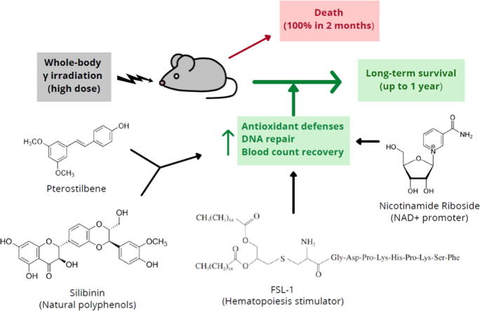
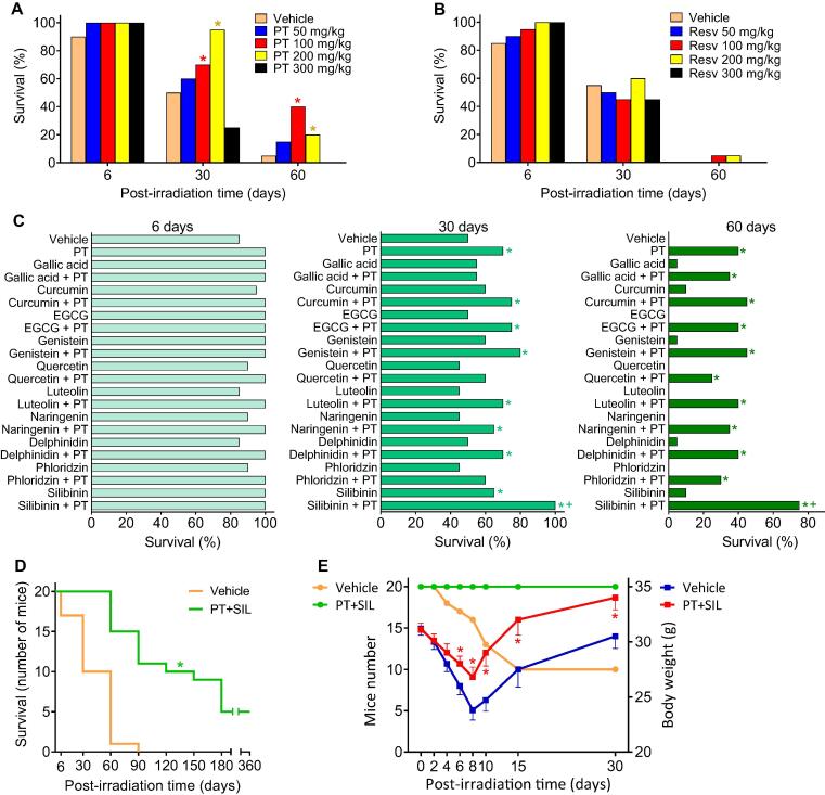
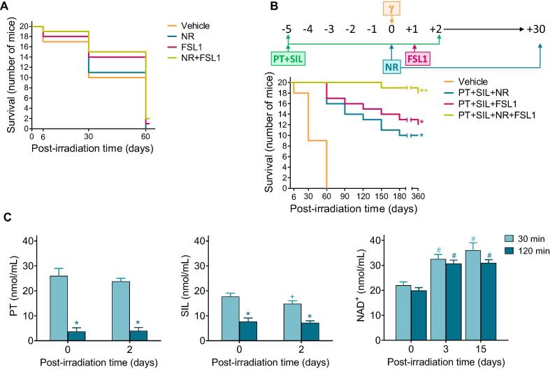
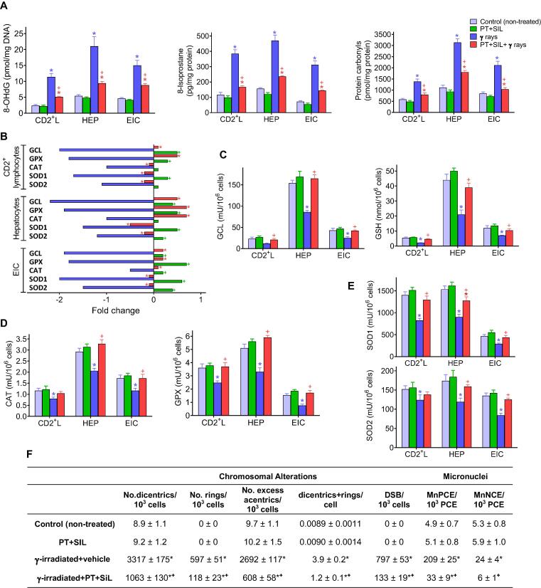
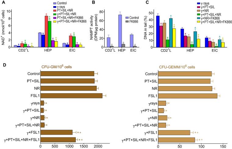
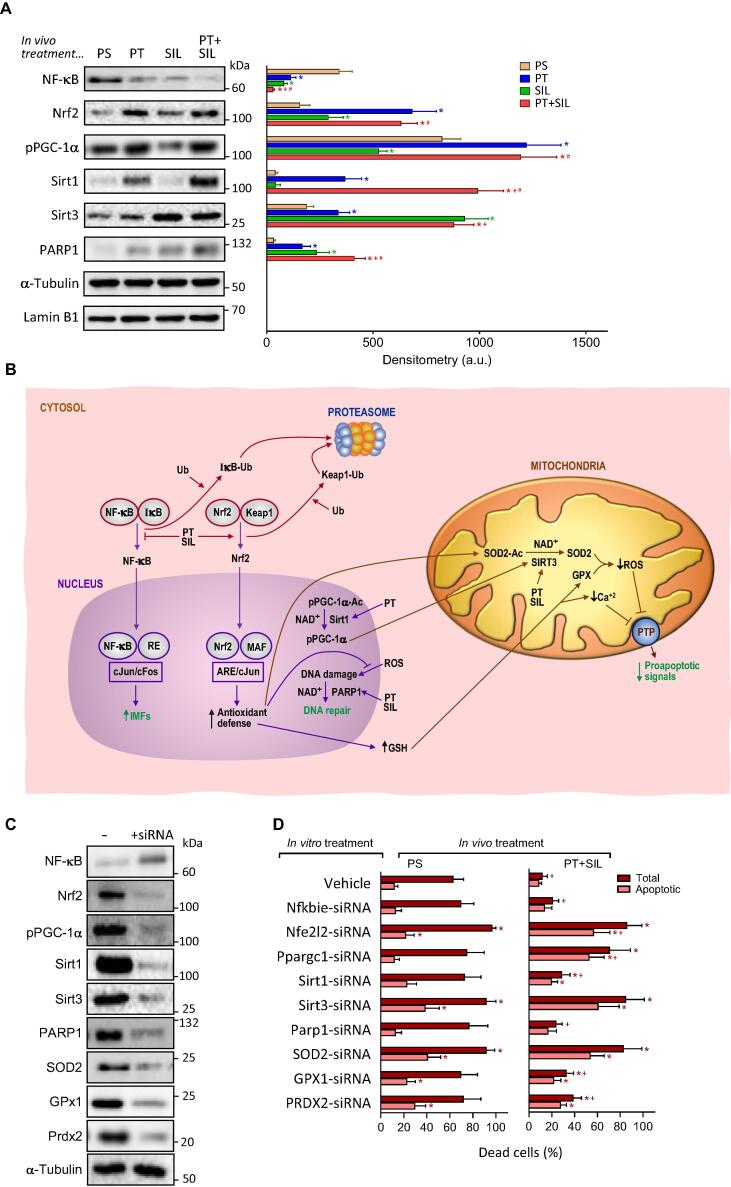
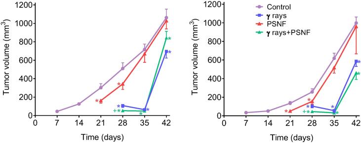
Results: The combination of pterostilbene (PT) and silibinin (SIL) was the most effective against γ-irradiation, resulting in 100% of the mice surviving at 30 days and 20% survival at one year. Treatment post γ-irradiation with two potential radiomitigators nicotinamide riboside (NR, a vitamin B3 derivative), and/or fibroblast-stimulating lipoprotein 1 (FSL1, a toll-like receptor 2/6 agonist), did not extend survival. However, the combination of PT, SIL, NR and FSL1 achieved a 90% survival one year post γ-irradiation. The mechanism involves induction of the Nrf2-dependent cellular antioxidant defense, reduction of NF-kB signaling, upregulation of the PGC-1α/sirtuins 1 and 3 axis, PARP1-dependent DNA repair, and stimulation of hematopoietic cell recovery. The pathway linking Nrf2, sirtuin 3 and SOD2 is key to radioprotection. Importantly, this combination did not interfere with X-ray mediated killing of different tumor cells in vivo.

Conclusion: The combination of the radioprotectors PT and SIL with the radiomitigators NR and FSL1 confer effective, long-term protection against γ radiation in vivo. This strategy is potentially capable of protecting mammals against ionizing radiations.

Keywords: Ionizing radiations; NAD(+) precursors; Natural polyphenols; Radioprotection; Toll-like receptor 2/6 ligands.


Conflict of interest statement

Declaration of Competing Interest The authors declare that they have no known competing financial interests or personal relationships that could have appeared to influence the work reported in this paper.

Figures

None
Graphical abstract
Fig. 1
Fig. 1
Effect of pterostilbene and silibinin on the survival of γ-irradiated mice. (A) Thirty-days survival of mice treated with γ rays (LD50/30) and PT. Mice (n = 20/group) were treated with PT (dissolved in 2-hydroxypropyl-b-cyclodextrin, then suspended in 0.3% carboxymethylcellulose; administered orally, once per day, 5 days before and 2 days after irradiation). Mice received whole-body γ radiation (one single dose of 7.2 Gy (137Cs) at 0.5–0.6 Gy/min). Data were analyzed with LogRank (Mantel-Cox) test (*p < 0.05, comparing all groups vs. controls treated with vehicle). (B) Thirty-days survival of mice treated with γ rays (LD50/30) and resveratrol (Resv). Mice (n = 20/group) were treated with Resv (dissolved and administered exactly as PT). Mice received whole-body γ radiation as indicated for PT-treated mice. (C) Combination of PT and other natural polyphenols in the prevention of the lethality induced by γ radiation (LD50/30). Mice (n = 20/group) were treated with PT and/or other polyphenols. PT was solubilized and administered as above. All other polyphenolic phytochemicals, representing main flavonoids and non-flavonoids were solubilized and administered exactly as PT. Doses for each phytochemical were selected from the max non-toxic doses published. Data were analyzed with LogRank (Mantel-Cox) test (*p < 0.05, comparing all groups vs. controls treated with vehicle; +p < 0.05, comparing all groups vs. the group treated with PT alone). (D) Effect of the combination of PT and SIL on the long-term survival of mice subjected to whole-body γ irradiation (LD50/30). Mice (n = 20/group) were treated IP with PT (100 mg/kg) and SIL (70 mg/kg) × 7 days (5 days before and 2 days after γ irradiation). For IP administration, a PT salt (PT phosphate disodium salt) and SIL salt (SIL-C-2′,3-dihydrogen succinate, disodium salt) were dissolved in sterile vaccination-grade water. The IP administration was selected to facilitate the regular administration to a high number of mice. Data were analyzed with Kaplan-Meier curves and LogRank (Mantel-Cox) test (*< 0.05, comparing the PT + SIL-treated group vs. controls treated with vehicle). (E) Survival of γ-irradiated (LD50/30) mice treated with PT and SIL and the effect on body weight. Mice (n = 20/group) were treated IP with PT and SIL (as in D). Statistical analyses (body weight data) were performed using Student’s t-test (*p < 0.01 comparing PT + SIL vs. vehicle-treated controls).
Fig. 2
Fig. 2
Combination of polyphenols with potential radiomitigators (nicotinamide riboside and fibroblast-stimulating lipopeptide 1) further increases the survival of γ-irradiated mice. (A) Effect of NR and FSL1 on the γ-irradiation (LD50/30)-induced mortality. Mice (n = 20/group) were treated with NR and/or FSL1. NR was dissolved in vaccination-grade water, and the pH of the solution neutralized (pH 7.0) before oral administration (185 mg/kg × day). FSL-1 was resuspended in sterile vaccination-grade water and administered IP (0.25 mg/kg × day). The volume administered in each case was of 50–60 mL depending on the weight of the mouse. NR was administered daily for 30 days, one single dose per day, and starting 1 h after the irradiation. FSL1 was administered only once, 24 h after the irradiation. Data were analyzed with LogRank (Mantel-Cox) test, but no significant differences were found comparing each treated-group vs. controls. (B) Effect of PT + SIL + NR + FSL1 on the survival of γ-irradiated mice (LD50/30). Mice (n = 20/group) were administered exactly as indicated in previous experiments [(A) and Fig. 1)]. Data were analyzed with Kaplan-Meier curves and LogRank (Mantel-Cox) test (*comparing all groups vs. vehicle-treated controls; +comparing the PT + SIL + NR + FSL1-treated group vs. the PT + SIL + NR- or the PT + SIL + FSL1-treated group). (C) PT, SIL and NAD+ levels in the circulating blood of treated and irradiated mice. Mice (n = 7/group) were treated with PT, SIL, or NR as scheduled previously and at the doses indicated in previous experiments (a). Statistical analyses were performed using Student’s t-test (p < 0.01; *comparing 120 min vs. 30 min; +comparing 2 vs. 0 days; #comparing 3 or 15 days vs. 0 days).
Fig. 3
Fig. 3
Protective effect of pterostilbene and silibinin on the γ-radiation-induced oxidative and cytogenetic damage. (A) Effect of PT + SIL on the levels of different oxidative damage-related molecular markers in cells (CD2+ lymphocytes, hepatocytes and epithelial intestinal) isolated from control and γ-irradiated mice. PT, SIL and γ rays were administered exactly as previously indicated in Fig. 2B (n = 5 per group, cell type, and treatment). Statistical analyses were performed using Student’s t-test (p < 0.01; *comparing all groups vs. controls; +comparing PT + SIL + γ rays-treated mice vs. mice treated with γ rays alone). (B) Expression (RT-PCR) of different antioxidant defense-related enzyme activities in isolated cells from PT + SIL- and/or γ-rays-treated mice. Fold change versus the expression rate of control (untreated) cells. PT, SIL and γ rays were administered as Fig. 2B (n = 5 per group, cell type, and treatment). Statistical analyses were performed using Student’s t-test (p < 0.01; *comparing PT + SIL + γ rays-treated mice vs. mice treated with γ rays alone; +comparing PT + SIL-treated mice vs. controls treated with vehicle). (C) Protective effect of PT + SIL on the γ radiation-induced damage in glutathione-related antioxidant defenses. PT, SIL and γ rays were administered as Fig. 2b (n = 5 per group, cell type, and treatment). Statistical analyses were performed using Student’s t-test (p < 0.01; *comparing all groups vs. controls; +comparing PT + SIL + γ rays-treated mice vs. mice treated with γ rays alone). (D) and (E) Protective effect of PT + SIL on the γ radiation-induced damage in CAT and GPX enzyme activities (D), and SOD enzyme activities (E). PT, SIL and γ rays were administered as Fig. 2B (n = 5 per group, cell type, and treatment). Statistical analyses were performed using Student’s t-test (p < 0.01; *comparing all groups vs. controls; +comparing PT + SIL + γ rays-treated mice vs. mice treated with γ rays alone). (F) Effect of PT + SIL on the γ radiation-induced chromosomal aberrations and micronuclei formation in circulating CD2+ lymphocytes. Lymphocytes were isolated from mice and procedures were as described under Experimental Section. DSBs, double-strand DNA breaks. Micronucleated polychromatic (MnPCE) and normochromatic (MnNCE) cells. Statistical analyses were performed using Student’s t-test (p < 0.01; *comparing all groups vs. controls; +comparing PT + SIL + γ rays-treated mice vs. mice treated with γ rays alone).
Fig. 4
Fig. 4
Effect of radioprotectors and radiomitigators on cellular NAD+ content, DNA damage, and hematopoietic recovery in γ-irradiated mice. (A) Effect of PT, SIL, NR and the specific noncompetitive inhibitor of nicotinamide phosphoribosyltransferase (NAMPT) FK866 on the NAD+ content of cells isolated from γ-irradiated mice (n = 5 per group, cell type, and treatment). FK866 was administered in vivo (15 mg/kg, twice daily by IP injection, during 6 days and starting 3 days prior to irradiation, see Fig. 2B), and was also present in the cell cultures (100 nM) . Cells were isolated 72 h after irradiation. Statistical analyses were performed using Student’s t-test (p < 0.01; *comparing all groups vs. controls; +comparing PT + SIL + NR + γ rays-treated mice or PT + SIL + NR + FK866 + γ rays-treated mice vs. mice treated with γ rays alone). (B) Effect of FK866 on the cellular NAMPT activity (p < 0.01; *comparing FK866-treated mice vs. controls, n = 5). Mice were treated with the inhibitor for 6 days as above (A). (C) Effect of PT, SIL, NR and FK866 on the DNA damage in cells isolated from γ-irradiated mice (n = 5 per group, cell type, and treatment). Statistical analyses were performed using Student’s t-test (p < 0.01; *comparing all groups vs. γ-irradiated controls; +comparing γ-irradiated groups where FK866 was present vs. their respective controls). Mice were treated and cells isolated as above (A). (D) Effect of PT, SIL, NR and FSL1 on the hematopoietic recovery in γ-irradiated mice. Hematopoietic colony forming cells CFU-GM and CFU-GEMM were quantified in femurs isolated from control and treated mice (n = 5 per group and treatment). Mice were treated as in Fig. 2B and bone marrow cells isolated 7 days after irradiation. Statistical analyses were performed using Student’s t-test (p < 0.01; *comparing all groups vs. controls; +comparing the different treatments + γ irradiation vs. γ irradiation alone).
Fig. 5
Fig. 5
Effect of the treatment with pterostilbene and silibinin on Nrf2-, NF-kB-, PARP1- and PGC1α-dependent mechanisms. Epithelial intestinal cells were isolated, right before the irradiation, from mice subjected to the protocol displayed in Fig. 2B. (A) Western blots for the detection of p65NF-kB, Nrf2, PARP1 and Sirt1 in nuclear extracts, Sirt3 in mitochondrial extracts, and phosphorylated PGC-1α in total extracts. Densitometric analysis (western blots) represents the mean values ± SD for 5–6 different mice per molecule and experimental condition (p < 0.01; *comparing PT-, SIL- or PT + SIL-treated mice versus physiological saline, PS-treated mice, +comparing PT + SIL- versus PT-treated mice; #comparing PT + SIL- versus SIL-treated mice). In vivo treatment (as in Fig. 2B) with PT and/or SIL and NR, FSL1 or NR + FSL1 did not alter significantly any of the data reported in the western blot analysis here displayed (results not shown). (B) Potential molecular interactions and interrelationships between different signaling mechanisms. Ub, ubiquitin; IkB, nuclear factor of kappa light polypeptide gene enhancer in B-cells inhibitor; Keap1, Kelch-like ECH-associated protein 1; RE, response element; ARE, antioxidant response element; MAF, musculoaponeurotic fibrosarcoma oncogene homolog; Ac, acetylated; IMFs, inflammation mediating factors. (C) Gene silencing of specific signaling-related molecules. The Fig. displays a representative experiment for n = 5–6 different experiments and experimental condition. PS, physiological saline. (D) Isolated IECs were cultured for 24 h in the presence or in the absence of specific siRNAs, and thereafter were irradiated with γ rays (same dose used for the in vivo experiments). Cell death was analyzed, as described under Methods, in isolated and cultured IEC cells 12 h after γ irradiation. PT (20 μM) and SIL (15 μM) were present in the cultures containing IECs isolated from PT + SIL-treated mice. Thereby, the effects induced by the in vivo treatment could be preserved under in vitro conditions. Data are mean values ± SD for 4–5 different experiments per molecule and experimental condition (p < 0.01; *comparing, under PS or PT + SIL, treatment with a specific siRNA versus controls -treatment with vehicle-; p < 0.01; +comparing data under PT + SIL versus their equivalents under PS).
Fig. 6
Fig. 6
Combined administration of the radioprotecting/radiomitigating combination (PT + SIL + NR + FSL-1; PSNF) and X rays to growing human A549 (lung adenocarcinoma) and MDA-MB-231 (triple negative mammary carcinoma) xenografts. Tumors were irradiated with X rays (10 Gy, single fraction, 5.0 Gy/min) using a 6-keV SL75 linear accelerator from Philips. The protocol for PSNF administration was identical to that followed in previous experiments (Fig. 2B), and the radiotherapy was administered on day 21. Statistical analyses (n = 5 mice per group) were performed using Student’s t-test (p < 0.01; *comparing all groups vs. controls; +comparing the X rays + PSNF-treated group vs. the group treated with X rays alone).

Similar articles

Cited by

References

    1. Riley P.A. Free radicals in biology: oxidative stress and the effects of ionizing radiation. Int J Radiat Biol. 1994;65:27–33. doi: 10.1080/09553009414550041. - DOI - PubMed
    1. Reisz J.A., Bansal N., Qian J., Zhao W., Furdui C.M. Effects of ionizing radiation on biological molecules–mechanisms of damage and emerging methods of detection. Antioxid Redox Signal. 2014;21:260–292. doi: 10.1089/ars.2013.5489. - DOI - PMC - PubMed
    1. Mavragani I.V., Laskaratou D.A., Frey B., Candéias S.M., Gaipl U.S., Lumniczky K., et al. Key mechanisms involved in ionizing radiation-induced systemic effects. A current review Toxicol Res (Camb) 2016;5:12–33. doi: 10.1039/c5tx00222b. - DOI - PMC - PubMed
    1. Patyar R.R., Patyar S. Role of drugs in the prevention and amelioration of radiation induced toxic effects. Eur J Pharmacol. 2018;819:207–216. doi: 10.1016/j.ejphar.2017.12.011. - DOI - PubMed
    1. Fischer N., Seo E.-J., Efferth T. Prevention from radiation damage by natural products. Phytomedicine. 2018;47:192–200. doi: 10.1016/j.phymed.2017.11.005. - DOI - PubMed

Publication types

LinkOut - more resources