

White matter changes should not exclude patients with idiopathic normal pressure hydrocephalus from shunt surgery
- PMID: 35599321
- PMCID: PMC9125842
- DOI: 10.1186/s12987-022-00338-8
White matter changes should not exclude patients with idiopathic normal pressure hydrocephalus from shunt surgery
Abstract
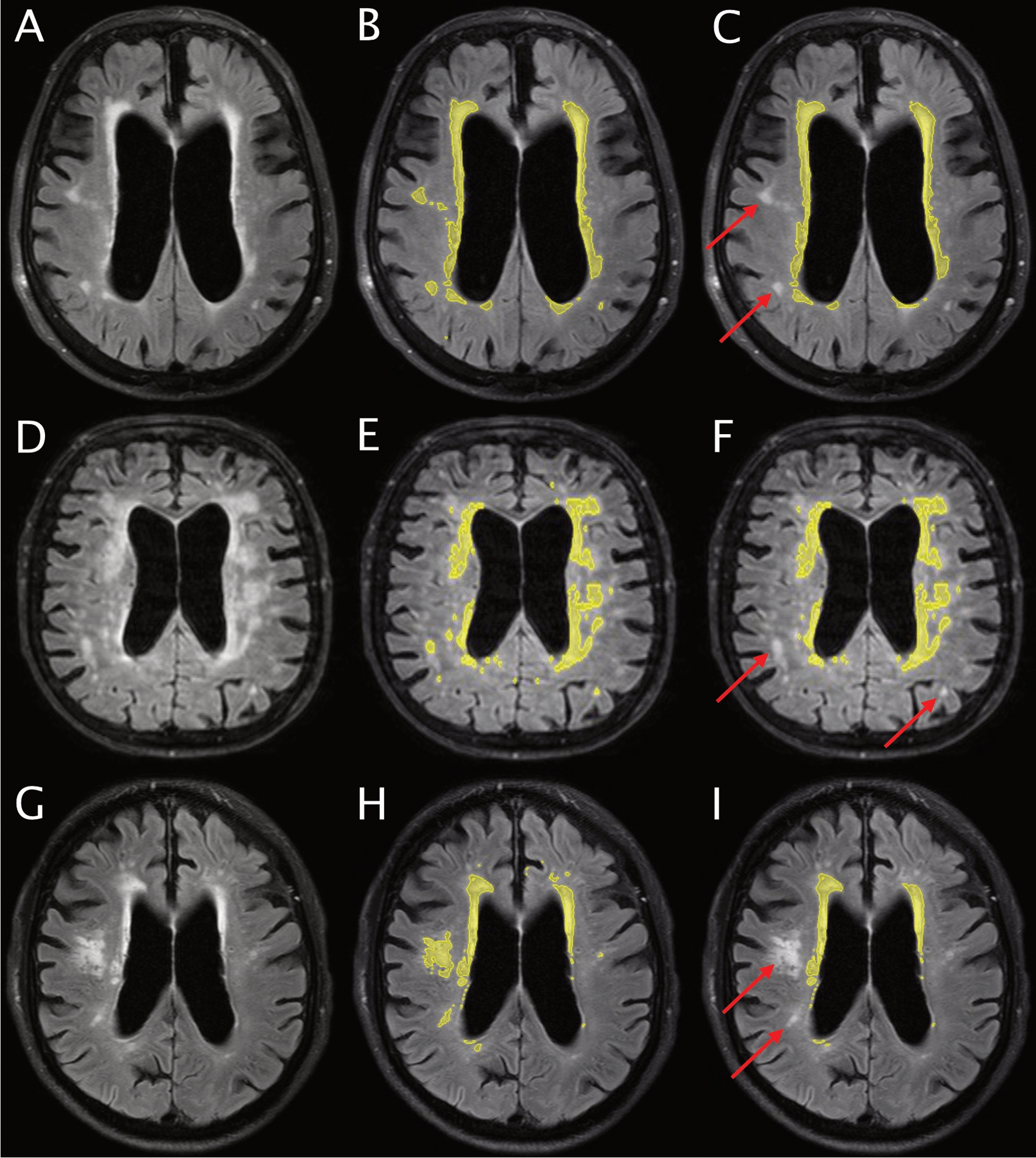
Introduction: White matter changes (WMC) on brain imaging can be classified as deep white matter hyperintensities (DWMH) or periventricular hyperintensities (PVH) and are frequently seen in patients with idiopathic normal pressure hydrocephalus (iNPH). Contradictory results have been reported on whether preoperative WMC are associated with outcome after shunt surgery in iNPH patients. The aim of this study was to investigate any association between DWMH and PVH and shunt outcome in patients with iNPH, using magnetic resonance volumetry.
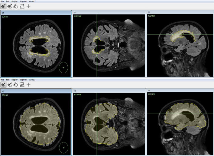
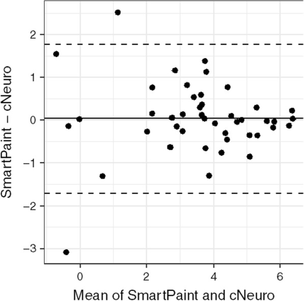
Methods: A total of 253 iNPH patients operated with shunt surgery and clinically assessed before and 12 months after surgery were included. All patients were investigated preoperatively with magnetic resonance imaging of the brain. The volumes of DWMH and PVH were quantified on fluid-attenuated inversion recovery images using an in-house semi-automatic volumetric segmentation software (SmartPaint). Shunt outcome was defined as the difference in symptom score between post- and preoperative investigations, measured on the iNPH scale, and shunt response was defined as improvement with ≥ 5 points.
Results: One year after shunt surgery, 51% of the patients were improved on the iNPH scale. When defining improvement as ≥ 5 points on the iNPH scale, there was no significant difference in preoperative volume of WMC between shunt responders and non-responders. If outcome was determined by a continuous variable, a larger volume of PVH was negatively associated with postoperative change in the total iNPH scale (p < 0.05) and negatively associated with improvement in gait (p < 0.01) after adjusting for age, sex, waiting time for surgery, preoperative level of symptoms, Evans' index, and disproportionately enlarged subarachnoid space hydrocephalus. The volume of DWMH was not associated with shunt outcome.
Conclusions: An association between outcome after shunt surgery and volume of PVH was seen, but there was no difference between shunt responders and non-responders in the volumes of DWMH and PVH. We conclude that preoperative assessment of WMC should not be used to exclude patients with iNPH from shunt surgery.
Keywords: Idiopathic normal pressure hydrocephalus; Magnetic resonance imaging; Shunt surgery outcome; Volumetric segmentation; White matter changes.
© 2022. The Author(s).
Conflict of interest statement
The authors declare that they have no competing interests.
Figures
References
-
- Jacobsson J, Qvarlander S, Eklund A, Malm J. Comparison of the CSF dynamics between patients with idiopathic normal pressure hydrocephalus and healthy volunteers. J Neurosurg. 2018;1–6. - PubMed
MeSH terms
LinkOut - more resources
Full Text Sources
