. 2022 May 19;6(4):e12718.
doi: 10.1002/rth2.12718. eCollection 2022 May.

Molecular analysis of vascular gene expression

Affiliations

Molecular analysis of vascular gene expression

Martijn A Van der Ent et al. Res Pract Thromb Haemost. .

Abstract

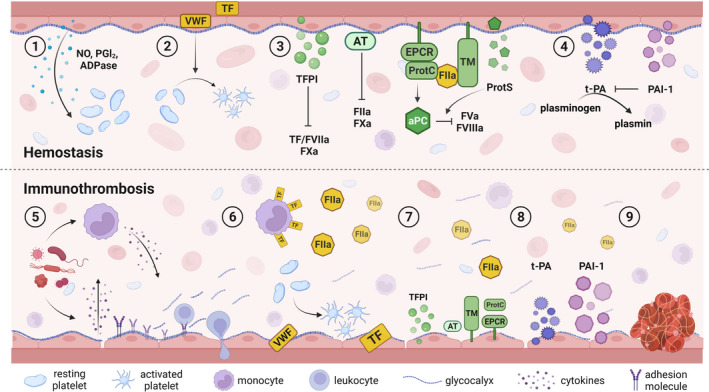
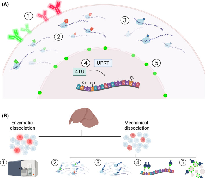
A State of the Art lecture entitled "Molecular Analysis of Vascular Gene Expression" was presented at the ISTH Congress in 2021. Endothelial cells (ECs) form a critical interface between the blood and underlying tissue environment, serving as a reactive barrier to maintain tissue homeostasis. ECs play an important role in not only coagulation, but also in the response to inflammation by connecting these two processes in the host defense against pathogens. Furthermore, ECs tailor their behavior to the needs of the microenvironment in which they reside, resulting in a broad display of EC phenotypes. While this heterogeneity has been acknowledged for decades, the contributing molecular mechanisms have only recently started to emerge due to technological advances. These include high-throughput sequencing combined with methods to isolate ECs directly from their native tissue environment, as well as sequencing samples at a high cellular resolution. In addition, the newest technologies simultaneously quantitate and visualize a multitude of RNA transcripts directly in tissue sections, thus providing spatial information. Understanding how ECs function in (patho)physiological conditions is crucial to develop new therapeutics as many diseases can directly affect the endothelium. Of particular relevance for thrombotic disorders, EC dysfunction can lead to a procoagulant, proinflammatory phenotype with increased vascular permeability that can result in coagulopathy and tissue damage, as seen in a number of infectious diseases, including sepsis and coronavirus disease 2019. In light of the current pandemic, we will summarize relevant new data on the latter topic presented during the 2021 ISTH Congress.

Keywords: coagulation; endothelial cells; gene expression; high‐throughput sequencing; inflammation.


Figures

FIGURE 1
FIGURE 1
Endothelial contributions to hemostasis and immunothrombosis. ECs play a role in hemostasis via expression of (1) factors that prevent platelet aggregation, (2) procoagulant VWF, which recruits and activates platelets upon vessel injury, (3) several anticoagulant factors that limit thrombin formation and (4) pro‐ and antifibrinolytic factors that are important for thrombus resolution. (5) Pathogen exposure activates immune cells and ECs, which stimulates cytokine release that induce expression of adhesion molecules leading to leukocyte recruitment and extravasation, shedding of the glycocalyx, and vascular leakage. Cytokines also (6) upregulate tissue factor expression on immune cells and activation of ECs leading to thrombin generation, while at the same time causing (7) a decrease in production of anticoagulant factors and (8) a shift toward inhibition of fibrinolysis, thereby resulting in (9) thrombus formation containing the pathogen. ADPase, adenophosphatase; aPC, activated protein C; AT, antithrombin; ECs, endothelial cells; EPCR, endothelial protein C receptor; FIIa, activated factor II; FVa, activated factor V; FVIIa, activated factor VII; FVIIIa, activated factor VIII; FXa, activated factor X; NO, nitric oxide; PAI‐1, plasminogen activator inhibitor 1; PGI2, prostacyclin; TF, tissue factor; TFPI, tissue factor pathway inhibitor; VWF, von Willebrand factor
FIGURE 2
FIGURE 2
Genetic approaches allowing cell type‐specific enrichment for molecular analyses. (A) In vivo labeling of cells can be achieved by (1) expression of a fluorescent surface protein, (2) tagging of a ribosomal protein with a fluorescent or (3) hemagglutin epitope tag, (4) TU tagging, or (5) via expression of a biotinylated nuclear envelope protein. (B) Ex vivo sample processing of (1) fluorescently labeled cells includes enzymatic dissociation followed by FACS sorting to isolate cells of interest. (2, 3) Tagged ribosomal proteins are incorporated into polysomal complexes, thereby enabling the isolation of actively translating mRNA from mechanically dissociated tissues via TRAP. (4) Administration of TU leads to production of thioRNA in cells that express UPRT. Nascent thioRNA is subsequently conjugated with biotin and isolated via streptavidin immunoprecipitation. (5) Streptavidin immunoprecipitation is also used to select nuclei containing a biotinylated nuclear envelope protein to evaluate epigenetic landscapes in a cell‐specific manner. FACS, fluorescence‐activated cell sorting; TRAP, translating ribosome affinity purification; TU, thiouracil; UPRT, uracil phosphoribosyltransferase

Similar articles

Cited by

References

    1. Aird WC. Phenotypic heterogeneity of the endothelium: I. Structure, function, and mechanisms. Circ Res. 2007;100:158‐173. - PubMed
    1. Augustin HG, Koh GY. Organotypic vasculature: from descriptive heterogeneity to functional pathophysiology. Science. 2017;357(6353):eaal2379. - PubMed
    1. Potente M, Makinen T. Vascular heterogeneity and specialization in development and disease. Nat Rev Mol Cell Biol. 2017;18:477‐494. - PubMed
    1. Mai J, Virtue A, Shen J, Wang H, Yang XF. An evolving new paradigm: endothelial cells–conditional innate immune cells. J Hematol Oncol. 2013;6:61. - PMC - PubMed
    1. Shao Y, Saredy J, Yang WY, et al. Vascular endothelial cells and innate immunity. Arterioscler Thromb Vasc Biol. 2020;40:e138‐e152. - PMC - PubMed