

Assessing the required glenoid peg penetration in native scapula when bone graft is used during primary and revision shoulder arthroplasty
- PMID: 35599713
- PMCID: PMC9121290
- DOI: 10.1177/1758573220987557
Assessing the required glenoid peg penetration in native scapula when bone graft is used during primary and revision shoulder arthroplasty
Abstract
Aims: Achieving purchase in native glenoid bone is essential for the stability of the glenoid baseplate when bone graft is used to address bone loss in both primary and revision shoulder arthroplasty procedures. The aim of this study is to assess the required depth of the baseplate peg in native bone when bone graft is used to result in satisfactory integration.
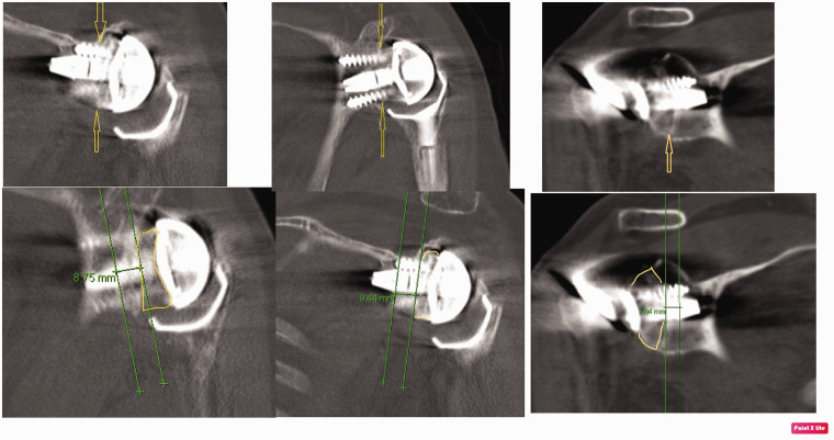
Patients and methods: The CT scans of patients who underwent either primary or revision arthroplasty procedures with bone graft using the SMR Axioma Trabecular Titanium (TT) Metal Backed glenoid system were assessed. We measured the depth of the glenoid peg in native glenoid bone. Measurements were taken by two authors separately.
Results: The scans of 53 patients (mean age 68 years) with a minimum follow-up of two years were reviewed. Implants included 12 anatomical and 41 reverse geometry prostheses. There were 17 primaries and 36 revisions: hemiarthroplasties (20) total (14) and reverse (2) implants. Bone grafts were from humeral head (15), iliac crest (34) and allograft (4). The mean depths were 8.8 mm (first assessor) and 9.10 mm (second assessor). The glenoid peg violated the glenoid vault in 32 patients and this did not adversely affect the outcome. There were three failures of implants all of which were aseptic failures and had peg penetration of less than 6 mm.
Conclusions: The mean depth of glenoid peg in native bone was 9 mm (variation between 0.2 and 0.52 mm at 95% confidence interval). Aseptic loosening was seen with peg penetration less than 6 mm in native bone. Glenoid vault violation was not associated with loosening.
Keywords: Glenoid peg depth; glenoid bone graft; native glenoid; primary shoulder arthroplasty; revision shoulder arthroplasty.
© 2021 The British Elbow & Shoulder Society.
Conflict of interest statement
Declaration of Conflicting Interests: The author(s) declared no potential conflicts of interest with respect to the research, authorship, and/or publication of this article.
Figures


Similar articles
-
Survivorship of autologous structural bone graft at a minimum of 2 years when used to address significant glenoid bone loss in primary and revision shoulder arthroplasty: a computed tomographic and clinical review.J Shoulder Elbow Surg. 2021 Mar;30(3):668-678. doi: 10.1016/j.jse.2020.06.015. Epub 2020 Jul 7. J Shoulder Elbow Surg. 2021. PMID: 32650067
-
Outcomes of femoral head allograft for the management of glenoid bone defects in revision reverse shoulder arthroplasty: a case-controlled study.J Shoulder Elbow Surg. 2023 Jun;32(6S):S32-S38. doi: 10.1016/j.jse.2022.12.022. Epub 2023 Jan 18. J Shoulder Elbow Surg. 2023. PMID: 36681105
-
Reconstruction of the glenoid using autologous bone-graft and the SMR Axioma TT metal-backed prosthesis: the first 45 sequential cases at a minimum of two years’ follow-up.Bone Joint J. 2018 Dec;100-B(12):1609-1617. doi: 10.1302/0301-620X.100B12.BJJ-2018-0494.R1. Bone Joint J. 2018. PMID: 30499322
-
[Glenoid reconstruction in revision shoulder arthroplasty].Oper Orthop Traumatol. 2019 Apr;31(2):98-114. doi: 10.1007/s00064-019-0594-8. Epub 2019 Mar 15. Oper Orthop Traumatol. 2019. PMID: 30874867 Review. German.
-
Aseptic Glenoid Baseplate Loosening After Reverse Total Shoulder Arthroplasty: A Systematic Review and Meta-Analysis.JBJS Rev. 2019 May;7(5):e7. doi: 10.2106/JBJS.RVW.18.00132. JBJS Rev. 2019. PMID: 31145263
References
-
- Day JS, Lau E, Ong KL, et al. Prevalence and projections of total shoulder and elbow arthroplasty in the United States to 2015. J Shoulder Elbow Surg 2010; 19: 1115–1120. - PubMed
-
- Klein SM, Dunning P, Mulieri P, et al. Effects of acquired glenoid bone defects on surgical technique and clinical outcomes in reverse shoulder arthroplasty. J Bone Joint Surg Am 2010; 92-A: 1144–1154. - PubMed
-
- Gunther SB, Lynch TL. Total shoulder replacement surgery with custom glenoid implants for severe bone deficiency. J Shoulder Elbow Surg 2012; 21: 675–684. - PubMed
-
- Walch G, Badet R, Boulahia A, et al. Morphologic study of the glenoid in primary glenohumeral osteoarthritis. J Arthroplasty 1999; 14: 756–760. - PubMed
-
- Page RS, Haines JF, Trail I. Impaction bone grafting of the glenoid in revision shoulder arthroplasty: classification, technical description and early results. Shoulder Elbow 2009; 1: 81–88.
LinkOut - more resources
Full Text Sources