

Shoulder electromyography activity during push-up variations: a scoping review
- PMID: 35599715
- PMCID: PMC9121296
- DOI: 10.1177/17585732211019373
Shoulder electromyography activity during push-up variations: a scoping review
Abstract
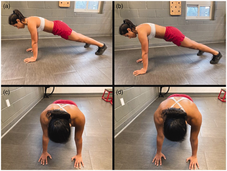
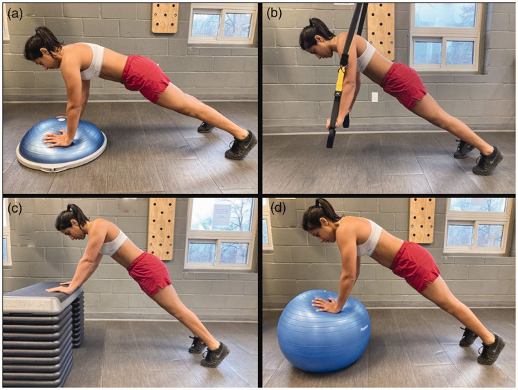
Background: Push-ups (PU) are a common closed chain exercise used to enhance shoulder girdle stability, with variations that alter the difficulty or target specific muscles. To appropriately select and prescribe PU exercises, an understanding of muscle activity during variations of the PU is needed. The purpose of this scoping review was to identify common PU variations and describe their muscle activation levels.
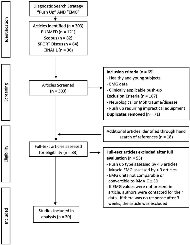
Methods: Databases searched included PubMed, CINAHL, Scopus, and SPORTDiscus for articles published between January 2000 and November 2019.
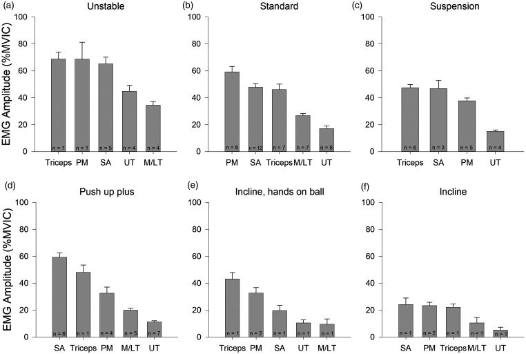
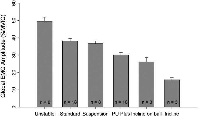
Results: Three hundred three articles were screened for eligibility with 30 articles included in the analysis. Six PU types and five muscles met the criteria for analysis. Weighted mean electromyography (EMG) amplitude was calculated for each muscle across PU types and for each PU type as a measure of global muscle activity. Triceps and pectoralis major had the highest EMG amplitude during unstable, suspension, incline with hands on a ball and the standard PU. Serratus anterior had the highest EMG amplitude during PU plus and incline PU. The greatest global EMG amplitude occurred during unstable surface PU.
Discussion: These results provide clinicians with a framework for prescribing PU to target specific muscles and scale exercise difficulty to facilitate rehabilitation outcomes.
Keywords: Push-up; electromyography; exercise; rehabilitation.
© 2021 The British Elbow & Shoulder Society.
Conflict of interest statement
Declaration of Conflicting Interests: The author(s) declared no potential conflicts of interest with respect to the research, authorship, and/or publication of this article.
Figures
References
-
- Reinold MM, Escamilla R, Wilk KE. Current concepts in the scientific and clinical rationale behind exercises for glenohumeral and scapulothoracic musculature. J Orthop Sports Phys Ther 2009; 39: 105–117. - PubMed
-
- Bleichert S, Renaud G, MacDermid J, et al. Rehabilitation of symptomatic atraumatic degenerative rotator cuff tears: a clinical commentary on assessment and management. J Hand Ther 2017; 30: 125–135. - PubMed
-
- Castelein B, Cagnie B, Cools A. Scapular muscle dysfunction associated with subacromial pain syndrome. J Hand Ther 2017; 30: 136–146. - PubMed
-
- Lephart SM, Pincivero DM, Giraldo JL, et al. The role of proprioception in the management and rehabilitation of athletic injuries. Am J Sports Med 1997; 25: 130–137. - PubMed
Publication types
LinkOut - more resources
Full Text Sources
Miscellaneous