Analysis of Therapeutic Targets of A Novel Peptide Athycaltide-1 in the Treatment of Isoproterenol-Induced Pathological Myocardial Hypertrophy
- PMID: 35599721
- PMCID: PMC9085328
- DOI: 10.1155/2022/2715084
Analysis of Therapeutic Targets of A Novel Peptide Athycaltide-1 in the Treatment of Isoproterenol-Induced Pathological Myocardial Hypertrophy
Abstract
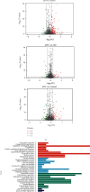
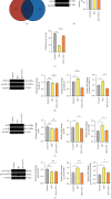
Myocardial hypertrophy is a pathological feature of many heart diseases. This is a complex process involving all types of cells in the heart and interactions with circulating cells. This study is aimed at identifying the differentially expressed proteins (DEPs) in myocardial hypertrophy rats induced by isoprenaline (ISO) and treated with novel peptide Athycaltide-1 (ATH-1) and exploring the mechanism of its improvement. ITRAQ was performed to compare the three different heart states in control group, ISO group, and ATH-1 group. Pairwise comparison showed that there were 121 DEPs in ISO/control (96 upregulated and 25 downregulated), 47 DEPs in ATH-1/ISO (27 upregulated and 20 downregulated), and 116 DEPs in ATH-1/control (77 upregulated and 39 downregulated). Protein network analysis was then performed using the STRING software. Functional analysis revealed that Hspa1 protein, oxidative stress, and MAPK signaling pathway were significantly involved in the occurrence and development of myocardial hypertrophy, which was further validated by vivo model. It is proved that ATH-1 can reduce the expression of Hspa1 protein and the level of oxidative stress in hypertrophic myocardium and further inhibit the phosphorylation of p38 MAPK, JNK, and ERK1/2.
Copyright © 2022 Xi Zheng et al.
Conflict of interest statement
Athycaltide-1 has obtained a granted patent (ZL201711418314.3) in China with Liying Hao, Jingyuan Li, Zhuo Li, Ze Kang and Siqi Wang as inventors. The authors have no other conflicts of interest.
Figures


Similar articles
-
The novel peptide athycaltide-1 attenuates Ang II-induced pathological myocardial hypertrophy by reducing ROS and inhibiting the activation of CaMKII and ERK1/2.Eur J Pharmacol. 2023 Oct 15;957:175969. doi: 10.1016/j.ejphar.2023.175969. Epub 2023 Aug 9. Eur J Pharmacol. 2023. PMID: 37567457
-
[Effects of fasudil on isoproterenol induced cardiac hypertrophy in rats].Zhongguo Ying Yong Sheng Li Xue Za Zhi. 2016 May 8;32(5):414-418. doi: 10.13459/j.cnki.cjap.2016.05.008. Zhongguo Ying Yong Sheng Li Xue Za Zhi. 2016. PMID: 29931844 Chinese.
-
Songling Xuemaikang Capsule inhibits isoproterenol-induced cardiac hypertrophy via CaMKIIδ and ERK1/2 pathways.J Ethnopharmacol. 2020 May 10;253:112660. doi: 10.1016/j.jep.2020.112660. Epub 2020 Feb 13. J Ethnopharmacol. 2020. PMID: 32061912
-
The CaMKII phosphorylation site Thr1604 in the CaV1.2 channel is involved in pathological myocardial hypertrophy in rats.Channels (Austin). 2020 Dec;14(1):151-162. doi: 10.1080/19336950.2020.1750189. Channels (Austin). 2020. PMID: 32290730 Free PMC article.
-
Effect of U50,488H, a κ-opioid receptor agonist on myocardial α-and β-myosin heavy chain expression and oxidative stress associated with isoproterenol-induced cardiac hypertrophy in rat.Mol Cell Biochem. 2010 Dec;345(1-2):231-40. doi: 10.1007/s11010-010-0577-4. Epub 2010 Aug 21. Mol Cell Biochem. 2010. PMID: 20730476
Cited by
-
Regulation of Cardiac Cav1.2 Channels by Calmodulin.Int J Mol Sci. 2023 Mar 29;24(7):6409. doi: 10.3390/ijms24076409. Int J Mol Sci. 2023. PMID: 37047381 Free PMC article. Review.
-
Mechanisms and Therapeutic Strategies for Myocardial Ischemia-Reperfusion Injury in Diabetic States.ACS Pharmacol Transl Sci. 2024 Nov 1;7(12):3691-3717. doi: 10.1021/acsptsci.4c00272. eCollection 2024 Dec 13. ACS Pharmacol Transl Sci. 2024. PMID: 39698288 Review.
References
-
- Fu Y., Xiao H., Zhang Y. Beta-adrenoceptor signaling pathways mediate cardiac pathological remodeling. Frontiers in Bioscience (Elite Edition) . 2012;4(5):1625–1637. - PubMed
MeSH terms
Substances
LinkOut - more resources
Full Text Sources
Research Materials
Miscellaneous

