. 2022 May 6:13:849571.
doi: 10.3389/fneur.2022.849571. eCollection 2022.

Progressive Brain Structural Impairment Assessed via Network and Causal Analysis in Patients With Hepatitis B Virus-Related Cirrhosis

Affiliations

Progressive Brain Structural Impairment Assessed via Network and Causal Analysis in Patients With Hepatitis B Virus-Related Cirrhosis

Shiwei Lin et al. Front Neurol. .

Abstract

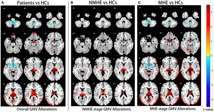
Objectives: This research amid to elucidate the disease stage-specific spatial patterns and the probable sequences of gray matter (GM) deterioration as well as the causal relationship among structural network components in hepatitis B virus-related cirrhosis (HBV-RC) patients.

Methods: Totally 30 HBV-RC patients and 38 healthy controls (HC) were recruited for this study. High-resolution T1-weighted magnetic resonance imaging and psychometric hepatic encephalopathy score (PHES) were evaluated in all participants. Voxel-based morphometry (VBM), structural covariance network (SCN), and causal SCN (CaSCN) were applied to identify the disease stage-specific GM abnormalities in morphology and network, as well as their causal relationship.

Results: Compared to HC (0.443 ± 0.073 cm3), the thalamus swelled significantly in the no minimal hepatic encephalopathy (NMHE) stage (0.607 ± 0.154 cm3, p <0.05, corrected) and further progressed and expanded to the bilateral basal ganglia, the cortices, and the cerebellum in the MHE stage (p < 0.05, corrected). Furthermore, the thalamus swelling had a causal effect on other parts of cortex-basal ganglia-thalamus circuits (p < 0.05, corrected), which was negatively correlated with cognitive performance (r = -0.422, p < 0.05). Moreover, the thalamus-related SCN also displayed progressive deterioration as the disease advanced in HBV-RC patients (p < 0.05, corrected).

Conclusion: Progressive deterioration of GM morphology and SCN exists in HBV-RC patients during advanced disease, displaying thalamus-related causal effects. These findings indicate that bilateral thalamus morphology as well as the thalamus-related network may serve as an in vivo biomarker for monitoring the progression of the disease in HBV-RC patients.

Keywords: MRI; cirrhosis; gray matter; hepatic encephalopathy; thalamus.


Conflict of interest statement

The authors declare that the research was conducted in the absence of any commercial or financial relationships that could be construed as a potential conflict of interest.

Figures

Figure 1
Figure 1
Study design schematic. (A) HBV-RC patients were categorized into NMHE and MHE according to PHES; overall and stage-specific gray matter (GM) alterations were calculated by comparing overall cirrhotic patients, NMHE patients, and MHE patients with HCs separately. (B) The bilateral thalamus was extracted as the seed region based on prior analysis for subsequent causal network of structural covariance (CaSCN) analysis and structural covariance network (SCN) analysis. (C) The thalamus-associated CaSCN was constructed, and sequenced alterations related to the thalamus were derived during the progression of MHE. (D) The correlation between the thalamus volume and PHES in HBV-RC patients was evaluated by Spearman's correlation analysis. (E) The thalamus-associated SCN was constructed, and stage-specific SCN alterations were calculated by a permutation test. GMV, gray matter volume; HC, healthy control; NMHE, patients without MHE; MHE, minimal hepatic encephalopathy; PHES, psychometric hepatic encephalopathy score; GCA, granger causality analysis; SCN, structural covariance network.
Figure 2
Figure 2
Overall and stage-specific gray matter alterations. Overall GM alterations in all cirrhotic patients mainly involved the cortex-basal ganglia-thalamus (CxBGTh) circuit and the thalamus–cerebellum circuit compared to HCs (A, false discovery rate [FDR] < 0.01). Meanwhile, stage-specific GM alteration started from the bilateral thalamus and the left caudate in the NMHE stage (compared with HCs, FDR < 0.05) (B), extending to other regions of the CxBGTh circuit and the thalamic-cerebellum circuit (including the bilateral basal ganglia, the bilateral cerebellum, and the cortex) in the MHE stage (compared with HCs, FDR < 0.05) (C). The t-values were represented by the colors shown in the color bar. GM, gray matter; HCs, healthy controls; NMHE, patients without MHE; MHE, minimal hepatic encephalopathy.
Figure 3
Figure 3
Thalamus volume differences among three groups. Thalamic volume displayed progressive swelling as the disease advanced in cirrhotic patients compared with HCs. HCs, healthy controls; NMHE, patients without MHE; MHE, minimal hepatic encephalopathy.
Figure 4
Figure 4
Voxel-based causal network of structural covariance (CaSCN). Thalamus swelling had causal effects on the left lingual gyrus, the left calcarine sulcus, Brodmann area 29, left middle occipital gyrus, left cuneus, left superior temporal gyrus, right Rolandic operculum, right frontal inferior operculum, right frontal middle gyrus, right frontal superior gyrus, left precentral gyrus, left paracentral lobule, bilateral postcentral gyrus, bilateral caudate nucleus, bilateral thalamus, and cerebellum (p < 0.05, corrected). Significant regions were overlaid onto the Montreal Neurological Institute (MNI) template, and the Granger causality value is represented by the colors shown in the color bar (A). The Sankey and Chord were also used to display the causal effects intuitively (B); the flow from the bilateral thalamus (red label) to the thalamus-affected regions (blue label) is colored in red according to the positive causal effects. GC, Granger causality; L, left; R, right; Sup, supper; Oper, opercularis; Mid, middle; Inf, inferior; Orb, orbitalis.
Figure 5
Figure 5
Spearman's correlations between the thalamus volume and PHES. The thalamus volume was negatively correlated with PHES performance in all cirrhosis patients. NMHE, patients without MHE; MHE, minimal hepatic encephalopathy; PHES, psychometric hepatic encephalopathy score.
Figure 6
Figure 6
Progressive deterioration of the thalamus-associated structural covariance network (SCN) in HBV-RC patients. Both HC and HBV-RC patients exhibited positive synchronous SCN between the thalamus and cortex (A,B). In HBV-RC patients, negative synchronous SCN between the thalamus and basal ganglia was also observed (B). The HBV-RC patients exhibited enhanced negative synchronous SCN between the thalamus and the bilateral basal ganglia as well as the cortex compared with HCs (C). Furthermore, progressive disruption of thalamus-related SCN was observed in HBV-RC patients as the disease advanced. Specifically, in the NMHE stage, patients showed increased negative synchronization between the basal ganglia and cortex compared to HCs (p < 0.01, corrected, D), and in the MHE stage, the negative synchronization between the thalamus and basal ganglia increased further compared to HCs (E) and NMHE patients (F). The color bar represents the t-values. HCs, healthy controls; NMHE, patients without MHE; MHE, minimal hepatic encephalopathy.

Similar articles

Cited by

References

    1. Wijdicks EF. Hepatic Encephalopathy. N Engl J Med. (2016) 375:1660–70. 10.1056/NEJMra1600561 - DOI - PubMed
    1. Tao R, Zhang J, You Z, Wei L, Fan Y, Cui J, et al. . The thalamus in cirrhotic patients with and without hepatic encephalopathy: a volumetric MRI study. Eur J Radiol. (2013) 82:e715–20. 10.1016/j.ejrad.2013.07.029 - DOI - PubMed
    1. Lu CQ, Jiao Y, Meng XP, Cai Y, Luan Y, Xu XM, et al. . Structural change of thalamus in cirrhotic patients with or without minimal hepatic encephalopathy and the relationship between thalamus volume and clinical indexes related to cirrhosis. Neuroimage Clin. (2018) 20:800–7. 10.1016/j.nicl.2018.09.015 - DOI - PMC - PubMed
    1. Butterworth R. Neuronal cell death in hepatic encephalopathy. Metab Brain Dis. (2007) 22:309–20. 10.1007/s11011-007-9072-3 - DOI - PubMed
    1. Seeley WW, Crawford RK, Zhou J, Miller BL, Greicius MD. Neurodegenerative diseases target large-scale human brain networks. Neuron. (2009) 62:42–52. 10.1016/j.neuron.2009.03.024 - DOI - PMC - PubMed

LinkOut - more resources