

Rhino-Orbital Cerebral Mucormycosis in a Patient With Diabetic Ketoacidosis: A Case Report and Literature Review
- PMID: 35599740
- PMCID: PMC9114505
- DOI: 10.3389/fneur.2022.815902
Rhino-Orbital Cerebral Mucormycosis in a Patient With Diabetic Ketoacidosis: A Case Report and Literature Review
Abstract
Background: Rhino-orbital cerebral mucormycosis (ROCM) is a rare, invasive, and fatal fungal disease. Due to the lack of specific clinical manifestations and adequate auxiliary examinations, patients are easily misdiagnosed in the early stage. Early diagnosis and timely therapy are essential for successful treatment.
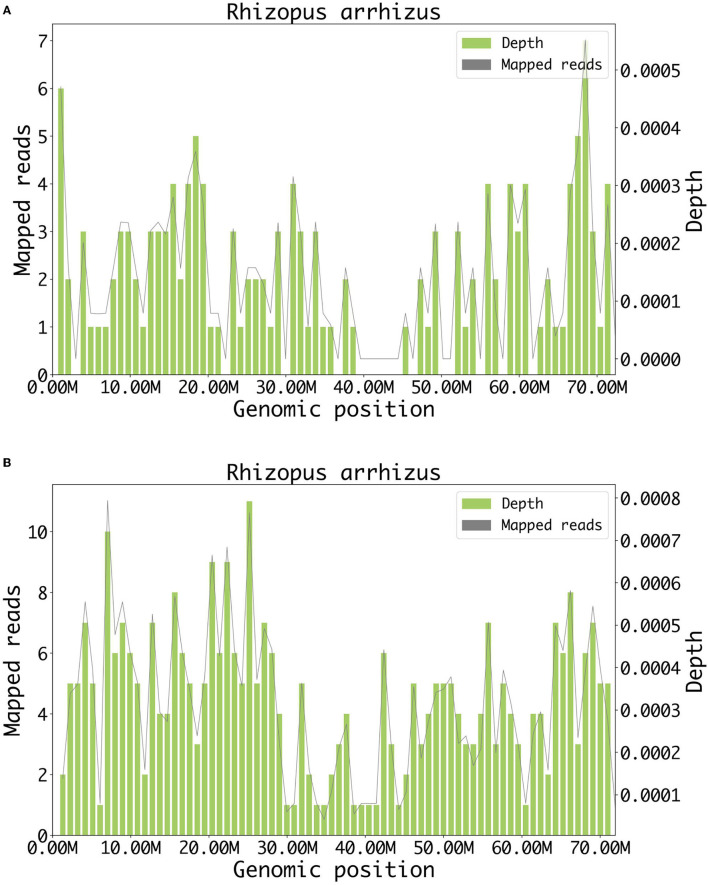
Case report: We report a 68-year-old man with diabetic ketoacidosis, presented with orbital apex syndrome (OAS), fever, and pansinusitis, which progressively worsened to death only 4 days after admission. It was finally confirmed as a fungal Rhizopus arrhizus infection by metagenomics cell-free DNA next-generation sequencing (mNGS) testing.
Conclusion: Orbital apex syndrome could be the initial presentation for mucormycosis. Thus, it is necessary to evaluate the presence of mucormycosis in patients with OAS, especially in diabetic or immunosuppressed hosts, and mNGS testing and timely antifungal therapy should be strongly recommended in highly suspected cases.
Keywords: diabetes; ketoacidosis; metagenomics cell-free DNA next-generation sequencing; orbital apex syndrome; rhino-orbital cerebral mucormycosis.
Copyright © 2022 Dong, Jordan, Shen, Wu, Guo, Zhao, Wang, Wang and Fang.
Conflict of interest statement
YW was employed by Genoxor Medical Science and Technology Inc. The remaining authors declare that the research was conducted in the absence of any commercial or financial relationships that could be construed as a potential conflict of interest.
Figures

References
Publication types
LinkOut - more resources
Full Text Sources
Miscellaneous