

In vitro maturation and in vivo stability of bioprinted human nasal cartilage
- PMID: 35599742
- PMCID: PMC9122109
- DOI: 10.1177/20417314221086368
In vitro maturation and in vivo stability of bioprinted human nasal cartilage
Abstract
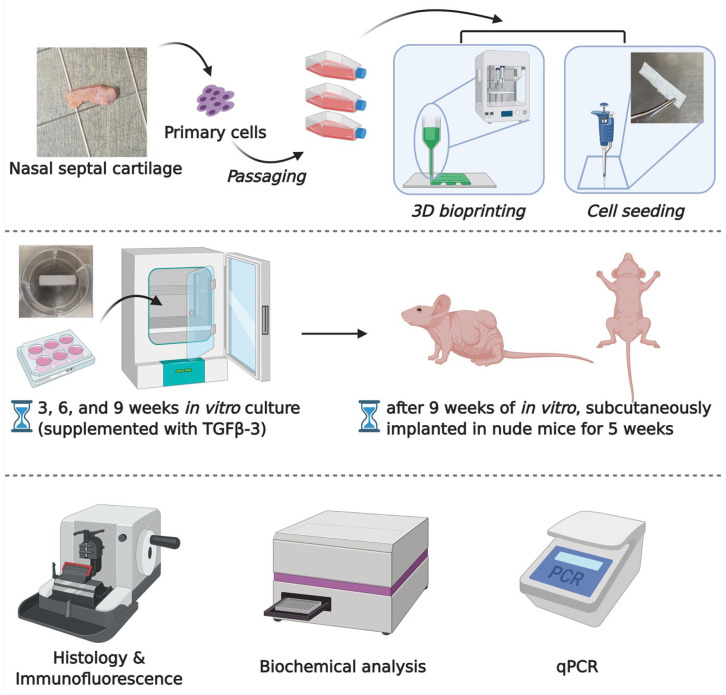
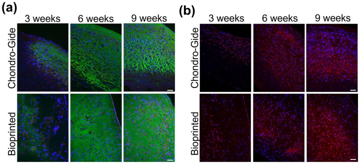
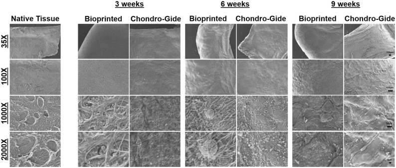
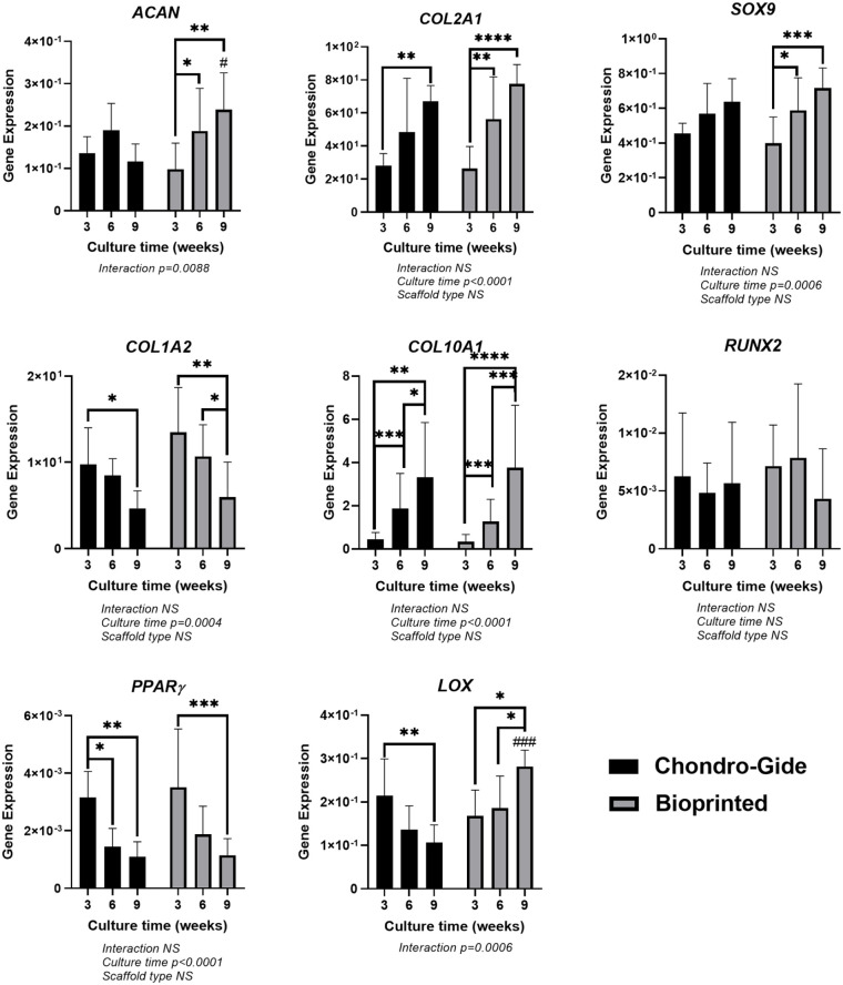
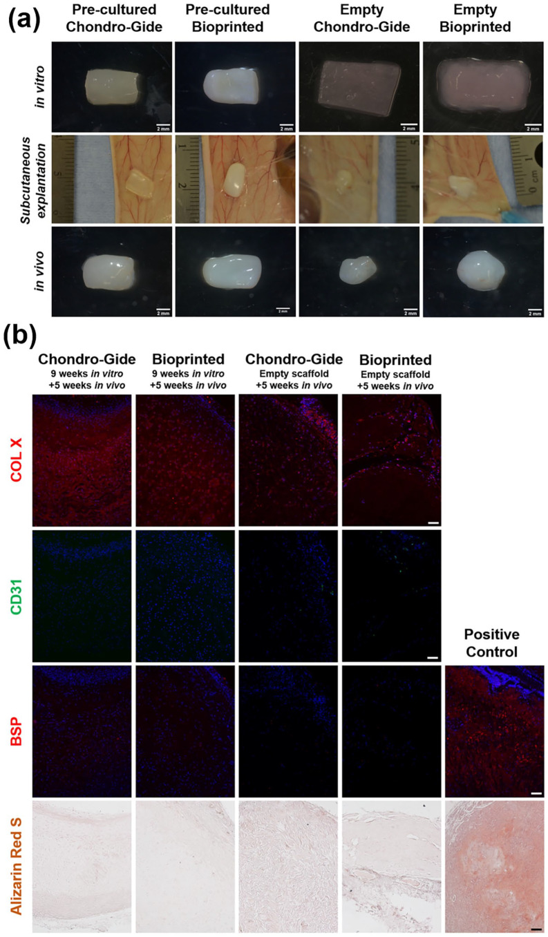
The removal of skin cancer lesions on the nose often results in the loss of nasal cartilage. The cartilage loss is either surgically replaced with autologous cartilage or synthetic grafts. However, these replacement options come with donor-site morbidity and resorption issues. 3-dimensional (3D) bioprinting technology offers the opportunity to engineer anatomical-shaped autologous nasal cartilage grafts. The 3D bioprinted cartilage grafts need to embody a mechanically competent extracellular matrix (ECM) to allow for surgical suturing and resistance to contraction during scar tissue formation. We investigated the effect of culture period on ECM formation and mechanical properties of 3D bioprinted constructs of human nasal chondrocytes (hNC)-laden type I collagen hydrogel in vitro and in vivo. Tissue-engineered nasal cartilage constructs developed from hNC culture in clinically approved collagen type I and type III semi-permeable membrane scaffold served as control. The resulting 3D bioprinted engineered nasal cartilage constructs were comparable or better than the controls both in vitro and in vivo. This study demonstrates that 3D bioprinted constructs of engineered nasal cartilage are feasible options in nasal cartilage reconstructive surgeries.
Keywords: Skin cancer; bioprinting; hydrogel; nasal cartilage; nude mice; septal chondrocytes; tissue engineering.
© The Author(s) 2022.
Conflict of interest statement
Declaration of conflicting interests: The author(s) declared no potential conflicts of interest with respect to the research, authorship, and/or publication of this article.
Figures


Similar articles
-
Multi-material 3D bioprinting of porous constructs for cartilage regeneration.Mater Sci Eng C Mater Biol Appl. 2020 Apr;109:110578. doi: 10.1016/j.msec.2019.110578. Epub 2019 Dec 20. Mater Sci Eng C Mater Biol Appl. 2020. PMID: 32228894
-
Bioprinting of human nasoseptal chondrocytes-laden collagen hydrogel for cartilage tissue engineering.FASEB J. 2021 Mar;35(3):e21191. doi: 10.1096/fj.202002081R. FASEB J. 2021. PMID: 33595884 Free PMC article.
-
Chondrocytes and stem cells in 3D-bioprinted structures create human cartilage in vivo.PLoS One. 2017 Dec 13;12(12):e0189428. doi: 10.1371/journal.pone.0189428. eCollection 2017. PLoS One. 2017. PMID: 29236765 Free PMC article.
-
Applications of 3D bioprinting in tissue engineering: advantages, deficiencies, improvements, and future perspectives.J Mater Chem B. 2021 Jul 14;9(27):5385-5413. doi: 10.1039/d1tb00172h. J Mater Chem B. 2021. PMID: 34124724 Review.
-
Toward tissue-engineering of nasal cartilages.Acta Biomater. 2019 Apr 1;88:42-56. doi: 10.1016/j.actbio.2019.02.025. Epub 2019 Feb 19. Acta Biomater. 2019. PMID: 30794988 Review.
Cited by
-
Recent advances in 3D bioprinted cartilage-mimicking constructs for applications in tissue engineering.Mater Today Bio. 2023 Nov 17;23:100870. doi: 10.1016/j.mtbio.2023.100870. eCollection 2023 Dec. Mater Today Bio. 2023. PMID: 38179226 Free PMC article. Review.
-
Biomaterials and tissue engineering strategies for posterior lamellar eyelid reconstruction: Replacement or regeneration?Bioeng Transl Med. 2023 Feb 15;8(4):e10497. doi: 10.1002/btm2.10497. eCollection 2023 Jul. Bioeng Transl Med. 2023. PMID: 37476060 Free PMC article. Review.
-
Head and Neck 3D Bioprinting-A Review on Recent Advancements in Soft Tissue 3D Bioprinting and Medical Applications.J Funct Biomater. 2025 Jun 30;16(7):240. doi: 10.3390/jfb16070240. J Funct Biomater. 2025. PMID: 40710454 Free PMC article. Review.
-
3D Bioprinting tissue analogs: Current development and translational implications.J Tissue Eng. 2023 Jul 13;14:20417314231187113. doi: 10.1177/20417314231187113. eCollection 2023 Jan-Dec. J Tissue Eng. 2023. PMID: 37464999 Free PMC article. Review.
-
A Contemporary Review of Trachea, Nose, and Ear Cartilage Bioengineering and Additive Manufacturing.Biomimetics (Basel). 2024 May 29;9(6):327. doi: 10.3390/biomimetics9060327. Biomimetics (Basel). 2024. PMID: 38921207 Free PMC article. Review.
References
-
- Madan V, Lear JT, Szeimies R-M. Non-melanoma skin cancer. Lancet 2010; 375: 673–685. - PubMed
-
- Lomas A, Leonardi-Bee J, Bath-Hextall F. A systematic review of worldwide incidence of nonmelanoma skin cancer. Br J Dermatol 2012; 166: 1069–1080. - PubMed
-
- Fulco I, Miot S, Haug MD, et al.. Engineered autologous cartilage tissue for nasal reconstruction after tumour resection: an observational first-in-human trial. Lancet 2014; 384: 337–346. - PubMed
-
- Burget GC, Menick FJ. The subunit principle in nasal reconstruction. Plast Reconstr Surg 1985; 76: 239–247. - PubMed
LinkOut - more resources
Full Text Sources
