. 2022 May 16:17:2191-2202.
doi: 10.2147/IJN.S358469. eCollection 2022.

Hydrophilic Realgar Nanocrystals Prolong the Survival of Refractory Acute Myeloid Leukemia Mice Through Inducing Multi-Lineage Differentiation and Apoptosis

Affiliations

Hydrophilic Realgar Nanocrystals Prolong the Survival of Refractory Acute Myeloid Leukemia Mice Through Inducing Multi-Lineage Differentiation and Apoptosis

Tao Wang et al. Int J Nanomedicine. .

Abstract

Introduction: Acute myeloid leukemia (AML) is a heterogeneous clonal disorder of hematopoietic progenitor cells, and the AML cells are differentiation retarded which results in the hyperproliferation of those malignant tumor cells. To stop the uncontrollable proliferation, inducing the AML cell differentiation is one highly expected therapy because it can bring relatively low systematic side effects compared to conventional chemotherapies; however, there are few options of inductive therapeutics in the clinical applications so far. This study aims to investigate the differentiation-induction effects of lab-developed hydrophilic nanocrystals of As4S4 (ee-As4S4).

Methods: In this work, ee-As4S4 was applied upon a refractory mouse model co-expressing AML1-ETO and HyC-KITD816V as well as a related human AML cell line, Kasumi-1, to investigate whether the nanocrystals can break the retardation of differentiation and drive the cells undergo apoptosis.

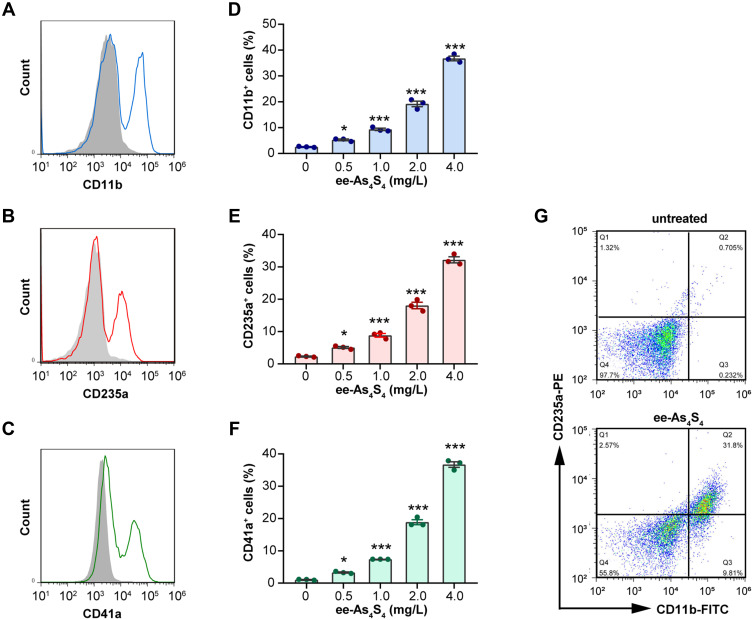
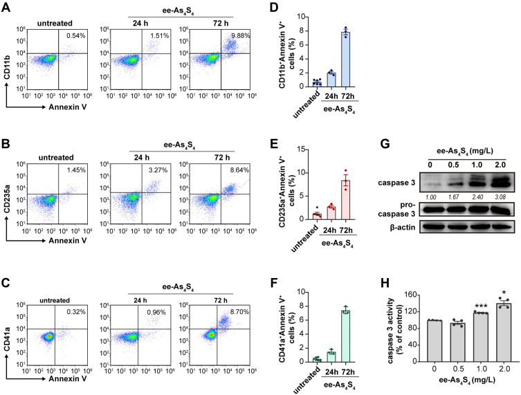
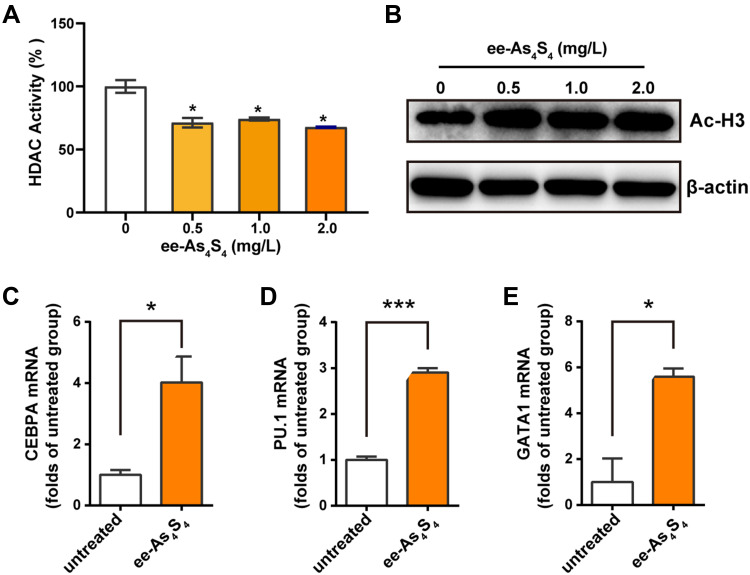
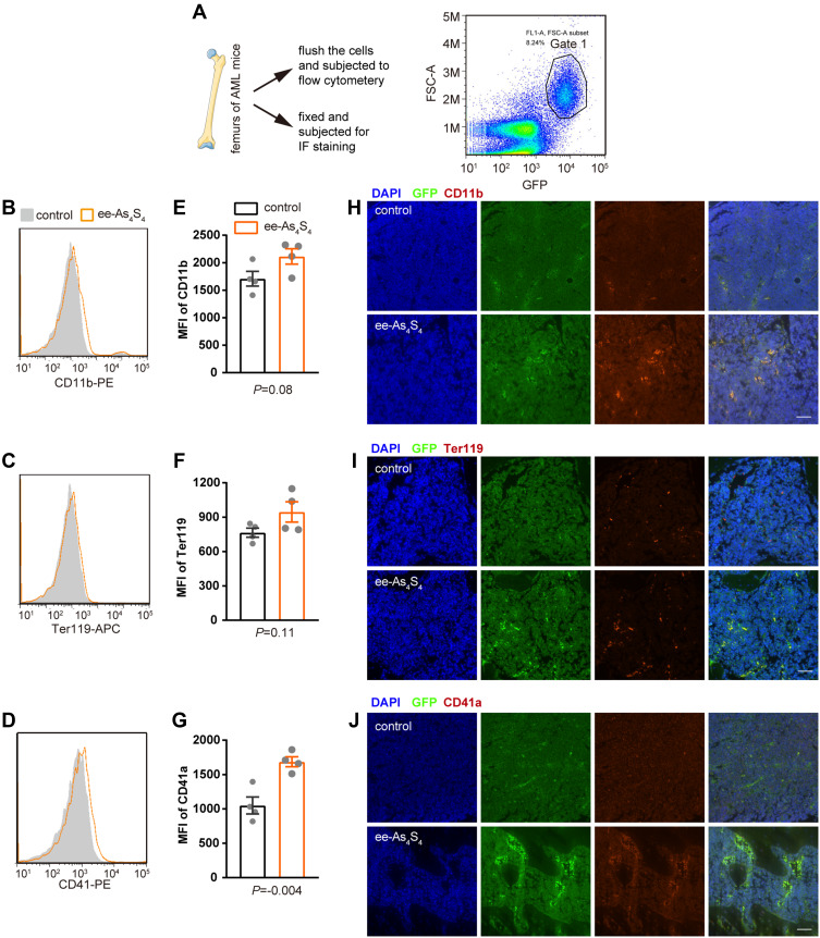
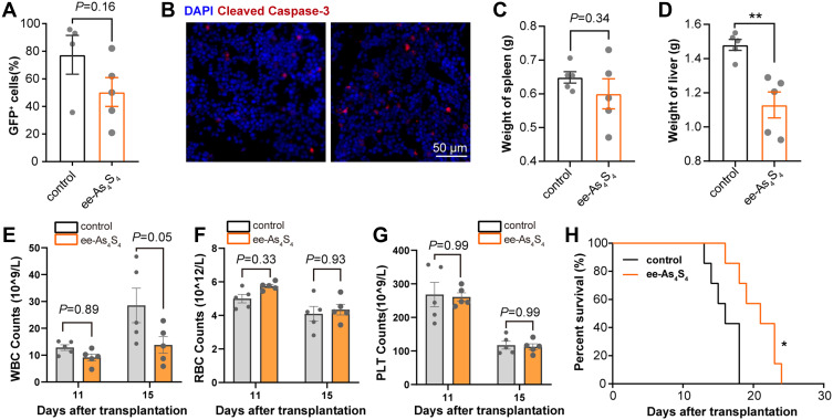
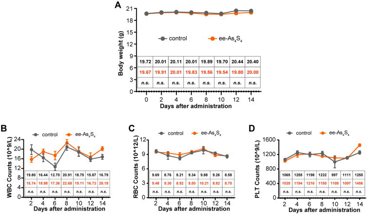
Results: It was shown that ee-As4S4 induced the upregulation of surface markers CD11b, CD235a, and CD41a, which indicate granulocytic, erythroid, and megakaryocytic differentiation respectively, leading to the multiple-lineage differentiation and post-differentiation apoptosis, and the inhibition of histone deacetylase activity was largely involved with the differentiation-induction effects. In the AML mice, orally administered ee-As4S4 increased the level of Ter119, CD11b, and CD41 in bone marrow-derived leukemia cells while reducing the percentage of leukemic cells in the bone marrow. Also, ee-As4S4 improved the hemogram and relieved the hepatomegaly and splenomegaly of the AML mice. As a result, the survival of the AML mice was significantly prolonged. Importantly, ee-As4S4 did not cause acute or chronic toxicity in healthy mice.

Conclusion: In conclusion, ee-As4S4 induced effective and multiple-lineage differentiation and apoptosis of AML cells in the refractory AML mouse model and cell line, suggesting that it holds promising potential as a novel inductive agent in differentiation therapy of AML.

Keywords: As4S4; acute myeloid leukemia; differentiation therapy; histone deacetylation; nanocrystals.


Conflict of interest statement

The authors declare no competing interest in this work.

Figures

Figure 1
Figure 1
The ee-As4S4 induced multi-lineage differentiation of Kasumi-1 cells. (AC) Overlap distribution of CD11b, CD235a, and CD41a. The filled gray peak indicated the control group, and the concentration of ee-As4S4 In (AC and G) is 4.0 mg/L. (DF) The portion of differentiation marker positive cells. (G) Plot distribution of Kasumi-1 cells defined by CD11b and CD235a antibodies. The incubation time for all experiments is 72 h. *P<0.05, ***P<0.001.
Figure 2
Figure 2
The ee-As4S4 induced post-differentiation apoptosis of Kasumi-1 cells. (AC) Representative plots distribution of the Kasumi-1 cells incubated with ee-As4S4 at 2.0 mg/L for 72 h after co-staining with Annexin V and antibodies against differentiation markers. (DF) Proportion of Annexin V and differentiation marker double-positive cells. (G) Western blot of pro-caspase 3 and caspase 3 after incubation with ee-As4S4 for 72 h. (H) Caspase-3 activity after incubation with ee-As4S4 for 72 h. *P<0.05, ***P<0.001.
Figure 3
Figure 3
Ee-As4S4 inhibited the activity of HDAC in Kasumi-1 cells. (A) HDAC activity in lysate Kasumi-1 cell that incubated with ee-As4S4 for 72 h. (B) The expression of acetylated histone H3 determined by Western Blotting in Kasumi-1 cell that incubated with ee-As4S4 for 72 h. (CE) Relative mRNA levels of CEBPA, PU.1, and GATA1 of Kasumi-1 cells incubated with ee-As4S4 at 2.0 mg/L for 48 h. *P<0.05 ***P<0.001.
Figure 4
Figure 4
The ee-As4S4 induced multi-lineage differentiation in vivo. (A) Illustration of the bone marrow assay and the gated scheme, cells in Gate 1 are leukemia cells. (BD) Overlap distribution of CD11b, Ter119, and CD41 expression in the leukemia cells in the control group and the ee-As4S4-treated group. (EG) Mean fluorescence intensity of the three markers in leukemia cells. (n=4) (HJ) Immunofluorescence staining of the three markers in femurs of mice from control and ee-As4S4 group.
Figure 5
Figure 5
The ee-As4S4 induced apoptosis in vivo. (A) The percentage of GFP+ leukemia cells in the bone marrow determined by flow cytometry. (B) Immunofluorescence staining of cleaved caspase-3 in the femur of mice from control and ee-As4S4 group. (C and D) The weight of spleen and liver of AML mice (n=5). (EG). The count of WBC, RBC, and PLT (n=5). (H) Survival curve of AML mice (n=7). *P < 0.05, **P<0.01.
Figure 6
Figure 6
The in vivo safety of ee-As4S4 in healthy mice. (A) The bodyweight of mice for each group (n=6). (BD) The counts of WBC (B), RBC (C), and PLT (D) were analyzed after the administration for 14 days (n=6). Mean of body weight, WBC, RBC, and PLT count were listed in the inserted table, the mean of the control group was black-colored, and that of the ee-As4S4 treated group is Orange-colored. “n.s.” indicates no significance between the two groups in the same day.

Similar articles

Cited by

References

    1. Bose P, Konopleva MY. ORY-1001: overcoming the differentiation block in AML. Cancer Cell. 2018;33(3):342–343. doi:10.1016/j.ccell.2018.02.014 - DOI - PubMed
    1. Dhall A, Zee BM, Yan F, Blanco MA. Intersection of epigenetic and metabolic regulation of histone modifications in acute myeloid leukemia. Front Oncol. 2019;9:432. doi:10.3389/fonc.2019.00432 - DOI - PMC - PubMed
    1. DiNardo CD, Cortes JE. Mutations in AML: prognostic and therapeutic implications. Hematology Am Soc Hematol Educ Program. 2016;2016(1):348–355. doi:10.1182/asheducation-2016.1.348 - DOI - PMC - PubMed
    1. Sykes DB, Kfoury YS, Mercier FE, et al. Inhibition of dihydroorotate dehydrogenase overcomes differentiation blockade in acute myeloid leukemia. Cell. 2016;167(1):171–186.e15. doi:10.1016/j.cell.2016.08.057 - DOI - PMC - PubMed
    1. de The H. Differentiation therapy revisited. Nat Rev Cancer. 2018;18(2):117–127. doi:10.1038/nrc.2017.103 - DOI - PubMed