. 2022 May 6:9:915818.
doi: 10.3389/fsurg.2022.915818. eCollection 2022.

Basal Cisternostomy for Severe TBI: Surgical Technique and Cadaveric Dissection

Affiliations

Basal Cisternostomy for Severe TBI: Surgical Technique and Cadaveric Dissection

L Giammattei et al. Front Surg. .

Erratum in

Abstract

Introduction: Cisternostomy is emerging as a novel surgical technique in the setting of severe brain trauma. Different surgical techniques have been proposed with a variable degree of epidural bone work. We present here the surgical technique as it is currently performed in our Institution.

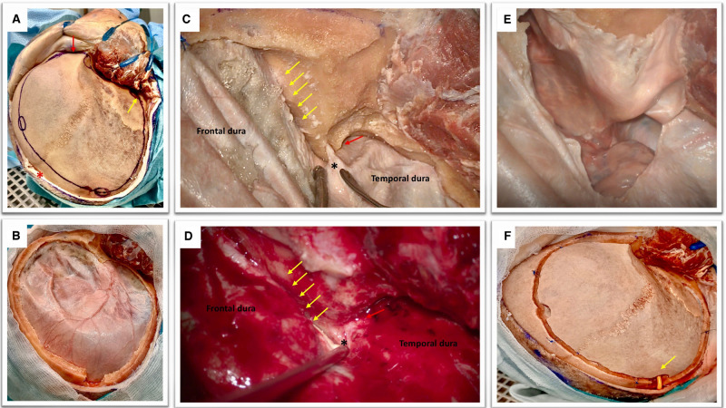
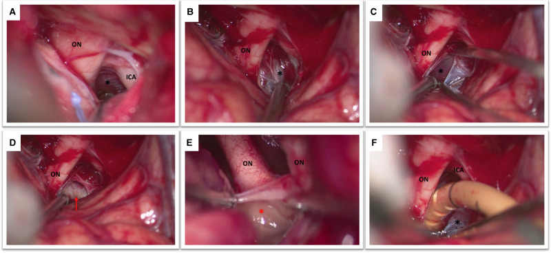
Methods: Anatomical dissection of one adult cadaveric head, injected and non-formalin fixed was perfomed. A large right fronto-temporo-parietal craniotomy was accomplished. Extradural sphenoidal drilling till opening of the superior orbital fissure was performed. The microsurgical anatomy of basal cisternostomy was then explored.

Results: A step by step description of the surgical technique, enriched with cadaveric and intraoperative images, was made.

Conclusion: Basal cisternostomy is a promising surgical technique that does not necessarily include complex surgical maneuvers. Trained neurosurgeon can safely implement it in their clinical practice.

Keywords: cadaveric dissection; cisternostomy; decompressive craniectomy (DC); severe brain trauma; skull base.


Conflict of interest statement

The authors declare that the research was conducted in the absence of any commercial or financial relationships that could be construed as a potential conflict of interest.

Figures

Figure 1
Figure 1
(A) Cadaveric dissection obtained after the elevation of soft tissues. The temporal muscle is retracted anteriorly with multiple hooks. The coronal suture is seen as well as the fontal, parietal, the squamous part of the temporal bones, the key hole and the orbital rim (red arrow). Please note the position of the midline (red asterisk) and the root of the zygoma (yellow arrow) that can be easily palpated in order to perform a craniotomy that is flush with the middle fossa. Note that the soft tissues are dissected till obtaining a visualization of the supra-orbital nerve and the orbital rim (red arrow).This is an essential step to perform a basal craniotomy that will enable the subsequent intradural elevation of the frontal lobe. Please note the position of the frontal and parietal burr holes that should be at a proper distance from the midline (approximately 2 cm) to avoid the risk of injury to the superior sagittal sinus. The key hole has an antero-superior location with respect to the pterion and should avoid transgression of the orbit with an appropriate direction of the perforator that should point in a posterior direction. (B) The obtained craniotomy allows an adequate fronto-basal access that would be increased by the epidural sphenoid drilling. The remaining part of the greater wing of the temporal bone is also drilled in order to expose the middle fossa floor and rapidly provide temporal lobe decompression. (C,D) Epidural sphenoidal drilling is accomplished by flattening the skull base to obtain a lateral subfrontal access (according to the direction of the yellow arrows) and continued till the opening of the superior orbital fissure (red arrow). The meningo-orbital band is identified (black asterisk); this is a dural band that tethers the frontotemporal dura to the orbit and is attached to the lateral part of the superior orbital fissure. The epidural drilling is usually stopped at this point. (E) A basal durotomy is performed and this small dural flap is antero-laterally retracted to increase the skull base view. (F) If the bone flap is replaced, care should be taken to ensure that the subcutaneously tunneled cisternal drain is not kinked (yellow arrow).
Figure 2
Figure 2
(A) This figure shows the view obtained after the opening of the optico-carotid cistern with optic nerve (ON), internal carotid artery (ICA) and Liliequist’s membrane (black asterisk). (B) The optic nerve (ON) and the diencephalic leaf of the Liliequist’s membrane (black asterisk) are seen. (C) The diencephalic leaf is opened enabling the exposure of the mesencephalic leaf (black asterisk). (D) The dorsum sellae (red arrow) becomes clearly visible (E) Both optic nerves and the lamina terminalis (red asterisk) are visible in this picture. (F) Note the position of the catheter which is placed in between the optic nerve (ON) and the internal carotid artery (ICA) passing through Liliequist’s membrane (black asterisk) into the prepontine cistern.

Similar articles

Cited by

References

    1. Peeters W, van den Brande R, Polinder S, Brazinova A, Steyerberg EW, Lingsma HF, et al. Epidemiology of traumatic brain injury in Europe. Acta Neurochir (Wien). (2015) 157(10):1683–96. 10.1007/s00701-015-2512-7 - DOI - PMC - PubMed
    1. Cherian I, Yi G, Munakomi S. Cisternostomy: replacing the age old decompressive hemicraniectomy? Asian J Neurosurg. (2013) 8(3):132–8. 10.4103/1793-5482.121684 - DOI - PMC - PubMed
    1. Giammattei L, Messerer M, Oddo M, Borsotti F, Levivier M, Daniel RT. Cisternostomy for refractory posttraumatic intracranial hypertension. World Neurosurg. (2018) 109:460–3. 10.1016/j.wneu.2017.10.085 - DOI - PubMed
    1. Giammattei L, Starnoni D, Maduri R, Bernini A, Abed-Maillard S, Rocca A, et al. Implementation of cisternostomy as adjuvant to decompressive craniectomy for the management of severe brain trauma. Acta Neurochir (Wien). (2020) 162(3):469–79. 10.1007/s00701-020-04222-y - DOI - PMC - PubMed
    1. Ramesh Chandra VV, Bodapati Chandra Mowliswara P, Banavath HN, Kalakoti CSR. Cisternostomy vs decompressive craniectomy for the management of traumatic brain injury: a randomized controlled trial. World Neurosurg. (2022) 19:S1878-8750(22)00214-5. 10.1016/j.wneu.2022.02.067 - DOI - PubMed

LinkOut - more resources