. 2022 May 4:13:891488.
doi: 10.3389/fpls.2022.891488. eCollection 2022.

Acquisition of Freezing Tolerance in Vaccinium macrocarpon Ait. Is a Multi-Factor Process Involving the Presence of an Ice Barrier at the Bud Base

Affiliations

Acquisition of Freezing Tolerance in Vaccinium macrocarpon Ait. Is a Multi-Factor Process Involving the Presence of an Ice Barrier at the Bud Base

Camilo Villouta et al. Front Plant Sci. .

Abstract

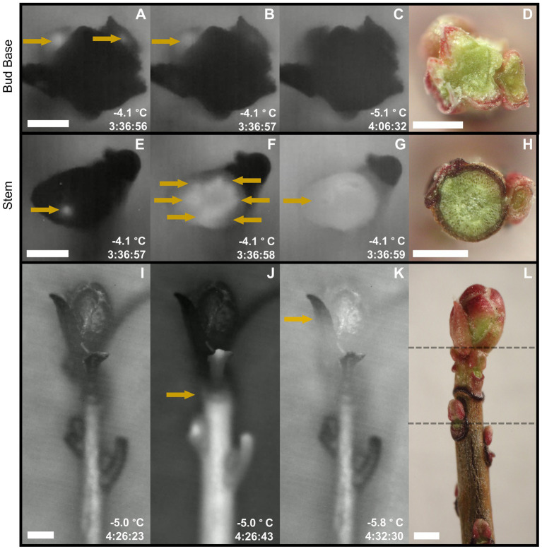
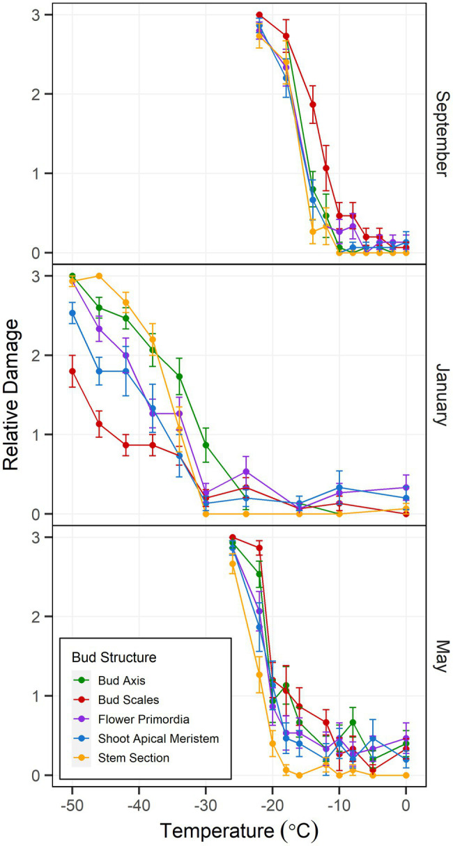
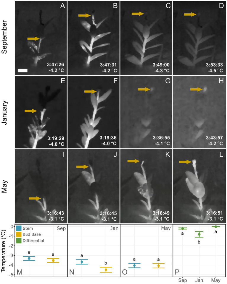
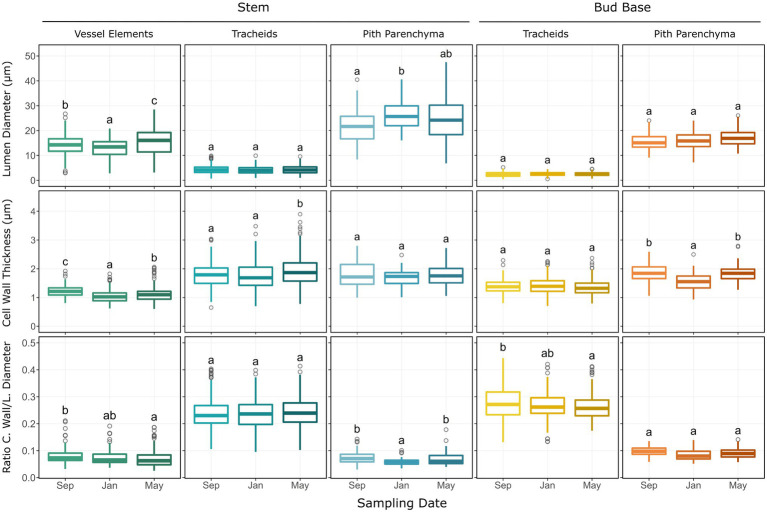
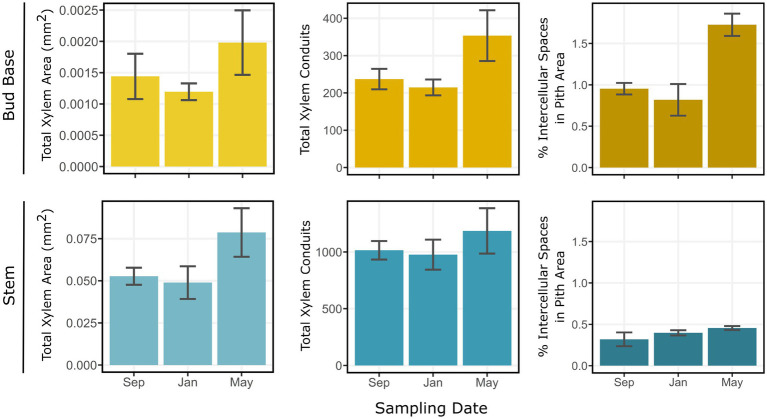
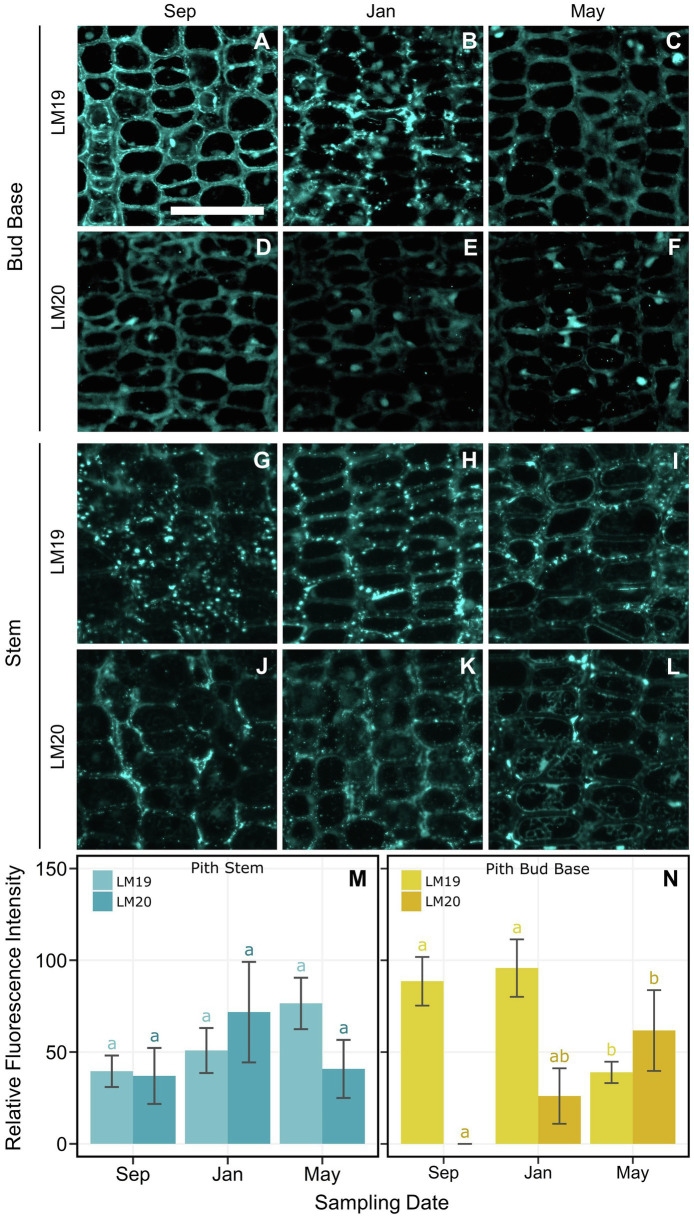
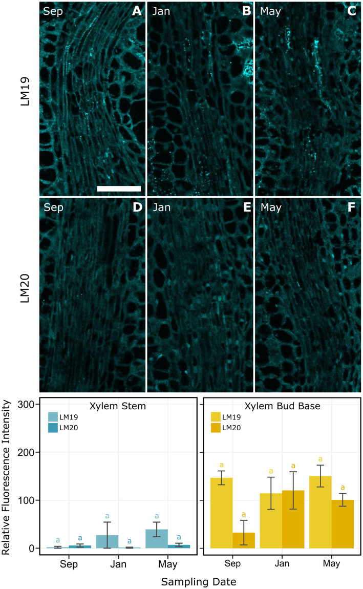
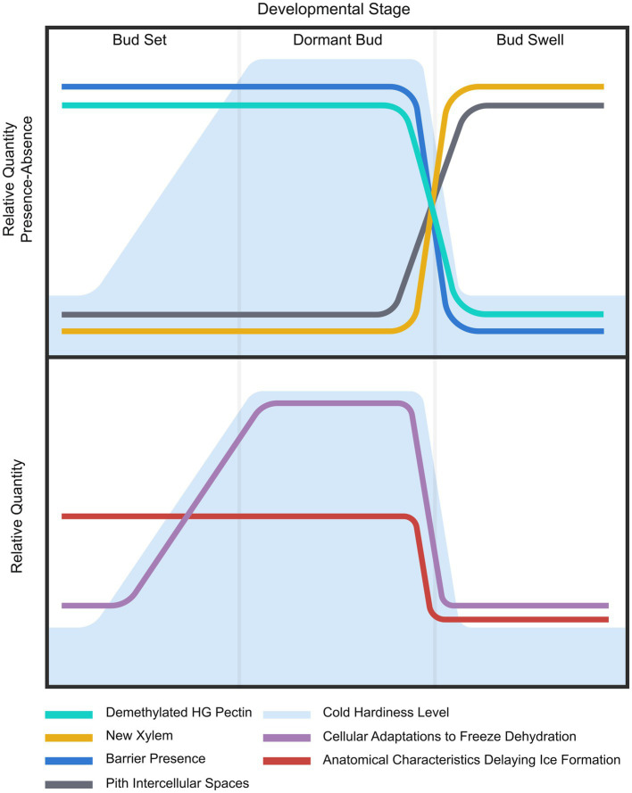
Bud freezing survival strategies have in common the presence of an ice barrier that impedes the propagation of lethally damaging ice from the stem into the internal structures of buds. Despite ice barriers' essential role in buds freezing stress survival, the nature of ice barriers in woody plants is not well understood. High-definition thermal recordings of Vaccinium macrocarpon Ait. buds explored the presence of an ice barrier at the bud base in September, January, and May. Light and confocal microscopy were used to evaluate the ice barrier region anatomy and cell wall composition related to their freezing tolerance. Buds had a temporal ice barrier at the bud base in September and January, although buds were only freezing tolerant in January. Lack of functionality of vascular tissues may contribute to the impedance of ice propagation. Pith tissue at the bud base had comparatively high levels of de-methyl-esterified homogalacturonan (HG), which may also block ice propagation. By May, the ice barrier was absent, xylogenesis had resumed, and de-methyl-esterified HG reached its lowest levels, translating into a loss of freezing tolerance. The structural components of the barrier had a constitutive nature, resulting in an asynchronous development of freezing tolerance between anatomical and metabolic adaptations.

Keywords: bud anatomy; cold acclimation; cold hardiness; cranberry (Vaccinium macrocarpon Ait); freeze dehydration; fruit crop; ice propagation.


Conflict of interest statement

The authors declare that the research was conducted in the absence of any commercial or financial relationships that could be construed as a potential conflict of interest.

Figures

Figure 1
Figure 1
Longitudinal and transversal high-definition thermal recordings of Vaccinium macrocarpon freezing patterns in freezing tolerant terminal buds. Time stamps represents the time elapse under the freezing treatment, only comparable under the same sampling date. Arrows highlight ice formation propagation. (A-C) Thermal images of transversal sections at the base of the bud. This sequence of images shows ice propagation from the base of the stem toward the exposed cut, where ice did not reach the surface. (D) Fresh cut of a transverse section made at the base of the bud. (E–G) Thermal images of transversal sections of the stem. This sequence of images shows ice propagation from the base of the stem toward the exposed cut, where ice reaches the surface. Ice formation reaches the surface first, most likely, through vascular tissues. (H) Fresh cut of a transversal section made at the stem zone. (I–K) Thermal images of shoots partially defoliated. This sequence of images shows ice propagation from the base of the stem toward the bud, where ice propagation stops at the base of the bud (J). Minutes later, ice formation is initiated in the bud (K). (L) Fresh shoot manually defoliated depicting location of bud base (upper dashed line) and stem zone (lower dashed line). Scale bar = 0.5 mm.
Figure 2
Figure 2
Controlled freezing test-incurred damage according to bud structure in Vaccinium macrocarpon terminal buds. Plant material was sampled in September 2018 and January and May 2019 from a commercial farm in Nekoosa, WI. Damage scoring was performed according to a discrete scale of damage from 0 to 3, where 0 represents no damage and 3 maximum damage. Damage was considered as the area with brown and water-soaked tissues. Each point was obtained from the average damage on each structure (n = 15). Vertical bars represent SE from the mean.
Figure 3
Figure 3
Thermal recordings of freezing patterns in Vaccinium macrocarpon shoots. Plant material was sampled in September 2018 and January and May 2019 from a commercial farm in Nekoosa, WI. Time stamps represents the time elapse under the freezing treatment, only comparable under the same structure. Arrows highlight ice formation propagation. (A–D) Ice propagation in a shoot sampled in September 2018. (B,C) Arrow indicates ice propagation stops at the base of the bud. (D) Shortly after, bud freezing resumes, indicated by the arrow. (E–H) Ice propagation in a shoot sampled in January 2019. (F,G) Arrow indicates where ice propagation stops at the base of the bud. (H) Minutes later, bud freezing resumes, signified by the arrow. (I–L) Ice propagation in a shoot sampled in May 2019. (J,K) Arrows indicate where ice propagation progresses through the stem. (G,H) Ice propagation progresses uninterrupted into the bud. (M–O) Average temperature of initiation of freezing in the stem or bud for (M) September 2018, (N) January 2019, and (O) May 2019. (P) Differences between the freezing temperature of stem and bud at each sampling date. Lower case letters represent significant difference among the means in each plot. Scale bar = 5 mm.
Figure 4
Figure 4
Images of transverse and longitudinal sections of the bud base and stem of Vaccinium macrocarpon shoots. Sections were stained with methylene FIGURE 4blue and basic fuchsin; dark blue indicates primary cell wall and pink/magenta secondary cell wall with high cellulose content. (A) Transverse section in the bud base and (B) in the stem zone both indicating xylem and pith area. (C–E) Transverse sections of vascular tissues in the bud base for (C) September, (D) January, and (E) May. (F–H) Transverse sections of pith tissue in the bud base for (F) September, (G) January, and (H) May. (I–K) Transverse sections of pith tissue in the stem for (I) September, (J) January, and (K) May. (L–N) Longitudinal sections of the bud base showing tracheids for (L) September, (M) January, and (N) May. X: Xylem, P: Pith. Scale bars (A,B) = 0.5 mm, (C–N) = 25 μm.
Figure 5
Figure 5
Histological parameters measured on transverse sections of Vaccinium macrocarpon terminal buds in the bud base and stem. Each plot depicts boxplots for each of the sampling dates. Each column represents a particular cell type or tissue, sub-grouped by stem and bud base. Rows represent the measured parameter. Whiskers represent the interquartile range 1.5-fold, while outliers are represented as gray rings. Lower case letters represent significant difference among the means in each plot.
Figure 6
Figure 6
Estimations for total xylem area (mm2), total xylem conduits, and percentage of intercellular spaces in the pith of Vaccinium macrocarpon terminal buds in the bud base and stem. Rows represent bud base and stem. Columns represent the estimated parameter. Vertical bars represent SE of the mean.
Figure 7
Figure 7
Indirect immunolocalization with antibodies (A–C,G–I) LM19 (de-methyl-esterified form) and (D–F,J–L) LM20 (methyl-esterified form), targeting homogalacturonan in pith cells of Vaccinium macrocarpon. (A–L) Columns represent sampling dates, rows represent sub-grouped evaluated zone, bud base or stem by antibody LM19 and LM20. (M) Bar plot for the average relative fluorescence intensity for pith cells in the stem. (N) Bar plot for the average relative fluorescence intensity for pith cells at the bud base. Lower case letters represent significant difference among the means of the same antibody and tissue in each plot. Vertical lines represent the SE of the mean. Scale bar = 50 μm.
Figure 8
Figure 8
Indirect immunolocalization with antibodies (A–C) LM19 (de-methyl-esterified form) and (D–F) LM20 (methyl-esterified form) targeting homogalacturonan in xylem and xylem parenchyma cells of Vaccinium macrocarpon. Images of samples from (A,D) September, (B,E) January, and (C,F) May. Bar plot for the average relative fluorescence intensity for xylem and xylem parenchyma cells in the stem zone (bottom left). Bar plot for the average relative fluorescence intensity for xylem and xylem parenchyma cells in the bud base (bottom right). Lower case letters represent significant difference among the means of the same antibody and tissue in each plot. Vertical lines represent the SE of the mean. Scale bar = 50 μm.
Figure 9
Figure 9
Summary of results and proposed hypothesis. (Upper) Lines depict general seasonal trends of evaluated elements considered part of the changes in cold hardiness in Vaccinium macrocarpon terminal buds. Background depicts variations in freezing tolerance. (Lower) Lines represent the proposed two levels of adaptations at anatomical and metabolic/cellular levels. Background depicts variations in freezing tolerance.

Similar articles

Cited by

References

    1. Addoms R., Mounce F. C. (1931). Notes on the nutrient requirements and the histology of the cranberry (Vaccinium macrocarpon Ait.) with special reference to mycorrhiza. Plant Physiol. 6, 653–668. doi: 10.1104/pp.6.4.653, PMID: - DOI - PMC - PubMed
    1. Aloni R. (2001). Foliar and axial aspects of vascular differentiation: hypotheses and evidence. J. Plant Growth Regul. 20, 22–34. doi: 10.1007/s003440010001 - DOI
    1. Aloni R. (2013). Role of hormones in controlling vascular differentiation and the mechanism of lateral root initiation. Planta 238, 819–830. doi: 10.1007/s00425-013-1927-8, PMID: - DOI - PubMed
    1. Andrews P. K., Proebsting E. L. (1986). Development of deep supercooling in acclimating sweet cherry and peach flower buds. HortScience 21, 99–100.
    1. Ashworth E. N. (1982). Properties of peach flower buds which facilitate supercooling. Plant Physiol. 70, 1475–1479. doi: 10.1104/pp.70.5.1475, PMID: - DOI - PMC - PubMed

LinkOut - more resources