. 2022 May 11:2022:6529842.
doi: 10.1155/2022/6529842. eCollection 2022.

Sulforaphane Ameliorates the Intestinal Injury in Necrotizing Enterocolitis by Regulating the PI3K/Akt/GSK-3 β Signaling Pathway

Affiliations

Sulforaphane Ameliorates the Intestinal Injury in Necrotizing Enterocolitis by Regulating the PI3K/Akt/GSK-3 β Signaling Pathway

Zhong-Kun Bao et al. Can J Gastroenterol Hepatol. .

Abstract

Objective: Necrotizing enterocolitis (NEC) is a serious neonatal disease; this study aims to investigate the role of sulforaphane (SFN) in NEC-induced intestinal injury.

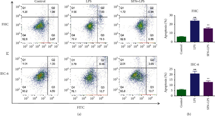
Methods: An animal model of NEC was established in newborn mice and intragastrically administrated with SFN; then, the general status and survival of the mice were observed. H&E staining was used to observe the pathological changes of intestinal tissues. ELISA, immunohistochemical staining, and flow cytometry assays were used to detect the levels of inflammatory factors, including TNF-α, IL-6, and IL-17, the expression of Bax, Bcl-2, TLR4, and NF-κB, and the percentages of the Th17 and Treg cells, respectively. GSK-3β expression levels were measured by immunofluorescence. IEC-6 and FHC cells were induced with LPS to mimic NEC in vitro and coincubated with SFN; then, the inflammatory factor levels and cell apoptosis rate were detected. Finally, Western blot was used to assess the expression of PI3K/Akt/GSK-3β pathway-related proteins in vitro and in vivo.

Results: SFN improved the survival rate of NEC mice during modeling, alleviated the severity of the intestinal injury, and reduced the proportion of Th17/Treg cells. SFN could inhibit TLR4 and NF-κB levels, decrease the release of inflammatory factors TNF-α and IL-6, suppress Bax expression, increase Bcl-2 expression, and inhibit apoptosis both in in vitro and in vivo models of NEC. Meanwhile, SFN regulated the expression of PI3K/Akt/GSK-3β pathway-related proteins in vitro and in vivo.

Conclusion: SFN relieved the inflammatory response and apoptosis by regulating the PI3K/Akt/GSK-3β signaling pathway, thereby alleviating NEC in model mice and cells.


Conflict of interest statement

The authors declare that they have no conflicts of interest.

Figures

Figure 1
Figure 1
Effects of SFN on survival rate and intestinal histopathological changes in NEC mice. (a) Survival analysis of mice in each group. (b) H&E staining and semiquantitative scoring results of the intestinal tissues of mice in each group, n = 6. NEC, necrotizing enterocolitis; SFN, sulforaphane. Compared with the control group, P < 0.05, ▲▲P < 0.01; compared with the NEC group, P < 0.05, ★★P < 0.01. Data represent the mean ± SD.
Figure 2
Figure 2
Changes of TLR4, MD-2, TNF-α, NF-κB, IL-6, CXCL1, and IL-17 contents in the serum of NEC mice. NEC, necrotizing enterocolitis; SFN, sulforaphane. Compared with the control group, P < 0.05, ▲▲P < 0.01; compared with the NEC group, P < 0.05, ★★P < 0.01. Data represent the mean ± SD, n = 6.
Figure 3
Figure 3
Changes in the proportion of Th17 and Treg cells in intestinal tissues of mice. NEC, necrotizing enterocolitis; SFN, sulforaphane. Compared with the control group, P < 0.05, ▲▲P < 0.01; compared with the NEC group, P < 0.05, ★★P < 0.01. Data represent the mean ± SD, n = 3.
Figure 4
Figure 4
Immunohistochemical staining of TLR4 (a), NF-κB (b), Bax (c), and Bcl-2 (d) and semiquantitative analysis of these proteins expression (e) in intestinal tissues of mice. NEC, necrotizing enterocolitis; SFN, sulforaphane. Compared with the control group, P < 0.05, ▲▲P < 0.01; compared with the NEC group, P < 0.05, ★★P < 0.01. Data represent the mean ± SD, n = 6.
Figure 5
Figure 5
Expression levels of PI3K/Akt/GSK-3β pathway-related proteins in intestinal tissue of NEC mice. Immunofluorescence staining results of GSK-3β (a) and fluorescence intensity (b) in intestinal tissues of mice in each group, n = 6, and expression of PI3K/Akt/GSK-3β pathway-related proteins in intestinal tissue of mice, n = 3. NEC, necrotizing enterocolitis; SFN, sulforaphane. Compared with the control group, P < 0.05, ▲▲P < 0.01; compared with the NEC group, P < 0.05, ★★P < 0.01. Data represent the mean ± SD.
Figure 6
Figure 6
The mRNA expression of TNF-α, IL-6, and IL-17 in FHC and IEC-6 cells. LPS, lipopolysaccharide; SFN, sulforaphane. Compared with the control group, P < 0.05, ▲▲P < 0.01; compared with the NEC group, P < 0.05, ★★P < 0.01. Data represent the mean ± SD, n = 3.
Figure 7
Figure 7
Apoptosis of FHC and IEC-6 cells. LPS, lipopolysaccharide; SFN, sulforaphane. Compared with the control group, P < 0.05, ▲▲P < 0.01; compared with the NEC group, P < 0.05, ★★P < 0.01. Data represent the mean ± SD, n = 3.
Figure 8
Figure 8
Expression of PI3K/Akt/GSK-3β pathway-related proteins in FHC (a) and IEC-6 (b) cells. LPS, lipopolysaccharide; SFN, sulforaphane. Compared with the control group, P < 0.05, ▲▲P < 0.01; compared with the NEC group, P < 0.05, ★★P < 0.01. Data represent the mean ± SD, n = 3.

References

    1. Eaton S., Rees C. M., Hall N. J. Current research on the epidemiology, pathogenesis, and management of necrotizing enterocolitis. Neonatology . 2017;111(4):423–430. doi: 10.1159/000458462. - DOI - PubMed
    1. Noor S., van de Graaf R. A., Rogier C. J., Reiss I. K. M., Vermeulen M. J. Risk factors for necrotizing enterocolitis in neonates: a systematic review of prognostic studies. BMC Pediatrics . 2017;17(1):p. 105. - PMC - PubMed
    1. Harpavat S., Pammi M., Gilger M. Novel treatments for NEC: keeping IBD in mind. Current Gastroenterology Reports . 2012;14(5):373–379. doi: 10.1007/s11894-012-0267-3. - DOI - PubMed
    1. Duan M., Han Z., Huang N. Changes of intestinal microflora in neonatal necrotizing enterocolitis: a single-center study. Journal of International Medical Research . 2020;48(9) doi: 10.1177/0300060520957804.300060520957804 - DOI - PMC - PubMed
    1. Yang K., Pagaling E., Yan T. Estimating the prevalence of potential enteropathogenic Escherichia coli and intimin gene diversity in a human community by monitoring sanitary sewage. Applied and Environmental Microbiology . 2014;80(1):119–127. doi: 10.1128/aem.02747-13. - DOI - PMC - PubMed