. 2022 Jun:48:101438.
doi: 10.1016/j.eclinm.2022.101438. Epub 2022 May 14.

Highly multiplexed immune repertoire sequencing links multiple lymphocyte classes with severity of response to COVID-19

Affiliations

Highly multiplexed immune repertoire sequencing links multiple lymphocyte classes with severity of response to COVID-19

Richard Dannebaum et al. EClinicalMedicine. 2022 Jun.

Abstract

Background: Disease progression of subjects with coronavirus disease 2019 (COVID-19) varies dramatically. Understanding the various types of immune response to SARS-CoV-2 is critical for better clinical management of coronavirus outbreaks and to potentially improve future therapies. Disease dynamics can be characterized by deciphering the adaptive immune response.

Methods: In this cross-sectional study we analyzed 117 peripheral blood immune repertoires from healthy controls and subjects with mild to severe COVID-19 disease to elucidate the interplay between B and T cells. We used an immune repertoire Primer Extension Target Enrichment method (immunoPETE) to sequence simultaneously human leukocyte antigen (HLA) restricted T cell receptor beta chain (TRB) and unrestricted T cell receptor delta chain (TRD) and immunoglobulin heavy chain (IgH) immune receptor repertoires. The distribution was analyzed of TRB, TRD and IgH clones between healthy and COVID-19 infected subjects. Using McFadden's Adjusted R2 variables were examined for a predictive model. The aim of this study is to analyze the influence of the adaptive immune repertoire on the severity of the disease (value on the World Health Organization Clinical Progression Scale) in COVID-19.

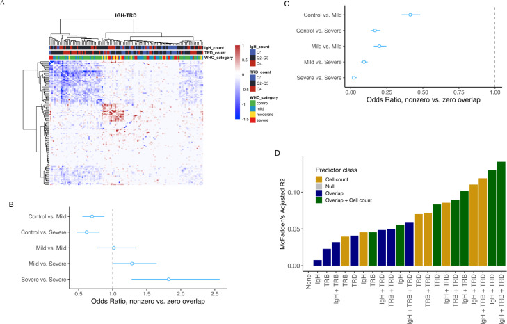
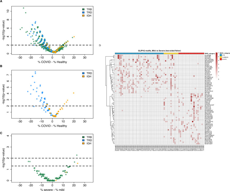
Findings: Combining clinical metadata with clonotypes of three immune receptor heavy chains (TRB, TRD, and IgH), we found significant associations between COVID-19 disease severity groups and immune receptor sequences of B and T cell compartments. Logistic regression showed an increase in shared IgH clonal types and decrease of TRD in subjects with severe COVID-19. The probability of finding shared clones of TRD clonal types was highest in healthy subjects (controls). Some specific TRB clones seems to be present in severe COVID-19 (Figure S7b). The most informative models (McFadden´s Adjusted R2=0.141) linked disease severity with immune repertoire measures across all three cell types, as well as receptor-specific cell counts, highlighting the importance of multiple lymphocyte classes in disease progression.

Interpretation: Adaptive immune receptor peripheral blood repertoire measures are associated with COVID-19 disease severity.

Funding: The study was funded with grants from the Berlin Institute of Health (BIH).

Keywords: COVID-19; Clinical course; Immune receptor; Immune repertoires.


Conflict of interest statement

Telman Dilduz, Dannenbaum Richard, Anja Blüher, Florian Rubelt, Gracie Du Zhipei, Luong Khai, Asgharin Hosseinali, Lin Hai and Berka Jan are employees of Roche Diagnostics and Dannenbaum Richard, Rubelt Forian, Lin Hai, Luong Khai, Berka Jan receive salary, stock and options as part of their employment compensation.

Figures

Figure 1
Figure 1
Cohort characteristics. (a) WHO score subject distribution of 3–8 by severity category: mild 3–4, moderate 5–6, and severe 7–8. (b) Donors age distribution for all samples used in the study, separated by healthy control and WHO severity groups. (c) The days since symptom onset, relative to sample collection timepoint. Boxplots were produced using the default settings in R. In all boxplots (Figure 1B and C), the bottom and top of the box are Q1 and Q3 (25th and 75th percentiles), and the black line near the middle of the box is Q2 (median, 50th percentile). The height of the box is the interquartile range (IQR=Q3Q1). The lower and upper whiskers are determined according to the following equations: Lowerwhisker=max(min(x),Q11.5×IQR)Upperwhisker=min(max(x),Q3+1.5×IQR) Where x is the variable being plotted.
Figure 2
Figure 2
Comparison of diversity metrics stratified by COVID-19 severity. (a) Cell count per ng input for PBMC samples. Severe samples have lower T/B-cell counts relative to mild and moderate (p-value 0.08312). (b) Cell count per ng input for whole blood samples. Severe samples have significantly lower cells counts relative to healthy samples (p-value 9.23E-06) and mild samples (p-value 0.007937). c) IgH per ng in PBMC samples. IgH concentrations were similar across all severity groups. (d) TRD per ng in PBMC samples, TRD concentrations were significantly lower in severe samples relative to mild (p-value 0.009778). (e) TRB per ng in PBMC samples. TRB concentrations were significantly lower in severe samples relative to mild (p-value 0.02562). (f) IgH Gini index by disease and severity group. IgH Gini index was significantly higher across all COVID-19 samples compared with healthy donors (p-value 4.334E-17) but consistent across mild, moderate, and severe groups. (g) TRD Gini index by disease and severity group. TRD Gini index was significantly higher across all COVID-19 samples compared with healthy donors (p-value 2.204E-4), but consistent across mild moderate, and severe groups. (h) TRB Gini index by disease and severity group. TRB Gini index was not significantly different between COVID-19 samples relative to healthy (p-value 0.1112), and not significantly different between the groups. . Boxplots were produced using the default settings in R. In all boxplots (Figure 2A–H), the bottom and top of the box are Q1 and Q3 (25th and 75th percentiles), and the black line near the middle of the box is Q2 (median, 50th percentile). The height of the box is the interquartile range (IQR=Q3Q1). The lower and upper whiskers are determined according to the following equations: Lowerwhisker=max(min(x),Q11.5×IQR)Upperwhisker=min(max(x),Q3+1.5×IQR) Where x is the variable being plotted.
Figure 3
Figure 3
Shared CDR3 in the IgH and TRD cell subsets differentiate mild and severe subjects. (a) IgH-TRD Jaccard overlap matrix. Both IgH and TRD matrices were median normalized against each other. Positive values represent a greater proportion of shared IgH clones and negative values represent a greater proportion of shared TRD clones. (b) Logistic regression was used to model the odds ratio of non-zero vs zero overlap between pairs of samples from the IgH Jaccard overlap matrix. The model comprised different COVID-19 severity groups, and compared them against a healthy-healthy baseline as indicated by the vertical dashed line. As disease severity increases, the odds of finding shared IgH clones also increases between COVID-19 individual pairs. (c) Logistic regression comparing the odds ratio of non-zero vs zero overlap between pairs of samples from the TRD Jaccard overlap matrix, for COVID-19 severity groups compared against healthy-healthy background. As disease severity increases, the odds of finding shared TRD clones decreases between COVID-19 individual pairs. (d) McFadden's adjusted pseudo-R for different models in terms of the association of COVID-19 clinical status (healthy, mild or severe) with sample covariates describing IgH-, TRB- and TRD- specific cell counts and clonal overlaps. In forest plots depicting logistic regression results (Figure 3B and C), the open circles are point estimates of odds ratios. The horizontal bars flanking them mark 95% confidence intervals of odds ratios.
Figure 4
Figure 4
CDR3 clonotypes across all 3 chains distinguish COVID-19 vs Healthy and Severe vs Mild. (a) Volcano plot of CDR3 clonotypes shared between the Healthy and COVID-19 subjects. Significance of overlap in the Healthy or COVID-19 cohorts was determined by randomization. For each clone,% shows the percentage of samples in the healthy or COVID-19 cohort in which that clone was detected (prevalence of the clone in that cohort). X axis shows the difference in the prevalence of each clone in the COVID-19 cohort and the healthy cohort. (b) Volcano plot comparing IgH and TRD enriched clonal types alone. TRD clonal types are disproportionately enriched in healthy samples while IgH clonal types are disproportionately enriched in COVID samples. (c) Analyzing COVID-19 enriched clones (p-value < 0.05) for differences between mild and severe, using Fisher's exact test. A few TRB clones were found to be significantly enriched in mild and severe samples. (d) Summary of GLIPH2 motifs identified, differentiating mild and severe samples. “%” indicates a position in the global pattern that allows amino acid variants.

References

    1. Huang C., Wang Y., Li X., Ren L., Zhao J., Hu Y., et al. Clinical features of patients infected with 2019 novel coronavirus in Wuhan, China. Lancet N Am Ed. 2020;395(10223):497–506. - PMC - PubMed
    1. Machhi J., Herskovitz J., Senan A.M., Dutta D., Nath B., Oleynikov M.D., et al. The natural history, pathobiology, and clinical manifestations of SARS-CoV-2 infections. J Neuroimmune Pharmacol. 2020:1–28. - PMC - PubMed
    1. Darab M.G., Keshavarz K., Sadeghi E., Shahmohamadi J., Kavosi Z. The economic burden of coronavirus disease 2019 (COVID-19): evidence from Iran. BMC Health Serv Res. 2021;21(1):1–7. - PMC - PubMed
    1. Guan W.J, Ni Z.Y, Hu Y., Liang W.H, Ou C.Q, He J.X, et al. Clinical characteristics of coronavirus disease 2019 in China. N Engl J Med. 2020;382(18):1708–1720. - PMC - PubMed
    1. Huang G., Kovalic A.J., Graber C.J. Prognostic value of leukocytosis and lymphopenia for coronavirus disease severity. Emerg Infect. Dis. 2020;26(8):1839. - PMC - PubMed