Review
. 2022 May 4:9:857581.
doi: 10.3389/fcvm.2022.857581. eCollection 2022.

Endocardial-Myocardial Interactions During Early Cardiac Differentiation and Trabeculation

Affiliations
Review

Endocardial-Myocardial Interactions During Early Cardiac Differentiation and Trabeculation

Xianghu Qu et al. Front Cardiovasc Med. .

Abstract

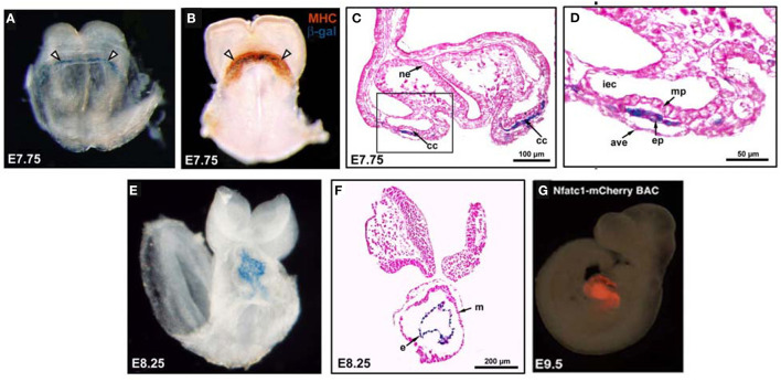
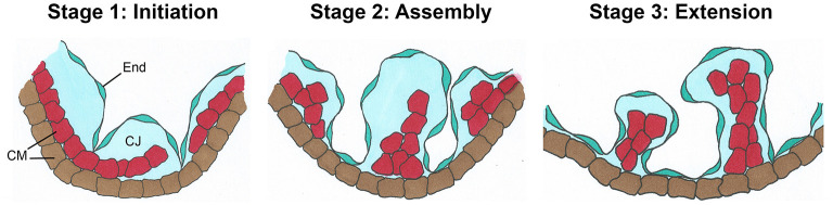
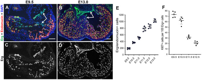
Throughout the continuum of heart formation, myocardial growth and differentiation occurs in concert with the development of a specialized population of endothelial cells lining the cardiac lumen, the endocardium. Once the endocardial cells are specified, they are in close juxtaposition to the cardiomyocytes, which facilitates communication between the two cell types that has been proven to be critical for both early cardiac development and later myocardial function. Endocardial cues orchestrate cardiomyocyte proliferation, survival, and organization. Additionally, the endocardium enables oxygenated blood to reach the cardiomyocytes. Cardiomyocytes, in turn, secrete factors that promote endocardial growth and function. As misregulation of this delicate and complex endocardial-myocardial interplay can result in congenital heart defects, further delineation of underlying genetic and molecular factors involved in cardiac paracrine signaling will be vital in the development of therapies to promote cardiac homeostasis and regeneration. Herein, we highlight the latest research that has advanced the elucidation of endocardial-myocardial interactions in early cardiac morphogenesis, including endocardial and myocardial crosstalk necessary for cellular differentiation and tissue remodeling during trabeculation, as well as signaling critical for endocardial growth during trabeculation.

Keywords: cardiac development; cardiomyocyte; endocardial cell; endocardial growth; endocardial-myocardial interactions; myocardial trabeculation.


Conflict of interest statement

The authors declare that the research was conducted in the absence of any commercial or financial relationships that could be construed as a potential conflict of interest.

Figures

Figure 1
Figure 1
The endocardium and myocardium are intimately associated during early differentiation. (A) An NFATc1-nuc-LacZ embryo, E7.75, stained with X-Gal. Nuclear expression of β-gal (blue) within the endocardium of the cardiac crescent (white arrowheads). (B) An E7.75 NFATc1-nuc-LacZ embryo stained with myosin heavy chain (MHC; reddish-brown) to identify the myocardium and co-stained with X-Gal to mark β-gal+ (blue) endocardium within the cardiac crescent (white arrowheads). (C,D) E7.75 embryo coronal sections revealing blue β-gal+ endocardial cells between the myocardial precursors and the anterior endoderm. mp, myocardial precursors; ep, endocardial precursor; ne, neuroectoderm; cc, cardiac crescent; ave, anterior visceral endoderm; iec, intraembryonic coelom. (E) X-Gal stained E8.25 NFATC1-nuc-LacZ transgenic embryo with blue β-gal expression confined to the endocardial layer of the linear heart tube. (F) Coronal sections of E8.25 NFATc1-nuc-LacZ embryo revealing blue β-gal expression specifically and exclusively in the endocardium. (G) Whole-mount immunofluorescence of an NFATc1-mCherry (BAC) E9.5 embryo confirms endocardium-specific mCherry expression [adapted from Misfeldt et al. (40) and Saint-Jean et al. (45)].
Figure 2
Figure 2
Schematic view of the three basic stages of trabeculation. Initiation (Stage 1) involves delamination of the innermost layer of cardiomyocytes (CMs, red) into the lumen where they form sheet-like protrusions called myocardial lamina. The endocardium (End) sends out angiogenic extensions, or sprouts, that penetrate the cardiac jelly (CJ) to directly touchdown onto the outer mycardial layer (brown). During Assembly, (Stage 2), endocardial sprouts first extend laterally underneath the myocardial lamina. Later they will assemble into individual short trabecular clusters within bubbles of cardiac jelly. Finally, in Stage 3, long sheet-like trabecular structures are formed by Extension [adapted from Qu et al. (72)].
Figure 3
Figure 3
Rapid expansion of the endocardium during trabeculation. (A–D) Dual immunostaining of wildtype mouse heart sections for the endothelial-specific ETS transcription factor Erg (green) and the endothelial marker endomucin (red) at E9.5 and E13.0. The endocardial cells in the ventricles display high expression of endomucin, but a subset of endocardial cells undergoing EndoMT to form the mesenchymal cells of the cardiac valves are negative for endomucin (arrows). Scale bars, 100 μm. (E) Quantification of the total number of endocardial cells (Erg+/endomucin+ cells) indicates a rapid expansion of endocardial cells from E9.5–E13.0 during trabeculation. (F) However, quantification of endocardial proliferation (Ki67/Erg-positive cells) after dual immunostaining of wildtype heart sections for Ki67 and Erg at E9.5–E12.5 shows a decrease in relative endocardial cell proliferation as trabeculation progresses.

References

    1. MacGrogan D, Münch J, de la Pompa JL. Notch and interacting signalling pathways in cardiac development, disease, and regeneration. Nat Rev Cardiol. (2018) 15:685–704. 10.1038/s41569-018-0100-2 - DOI - PubMed
    1. Buijtendijk MFJ, Barnett P, van den Hoff MJB. Development of the human heart. Am J Med Genet C Semin Med Genet. (2020) 184:7–22. 10.1002/ajmg.c.31778 - DOI - PMC - PubMed
    1. Brutsaert DL. Cardiac endothelial-myocardial signaling: its role in cardiac growth, contractile performance, and rhythmicity. Physiol Rev. (2003) 83:59–115. 10.1152/physrev.00017.2002 - DOI - PubMed
    1. Tian Y, Morrisey EE. Importance of myocyte-nonmyocyte interactions in cardiac development and disease. Circulation Res. (2012) 110:1023–34. 10.1161/CIRCRESAHA.111.243899 - DOI - PMC - PubMed
    1. Dye B, Lincoln J. The Endocardium and Heart Valves. Cold Spring Harb Perspect Biol. (2020) 12:a036723. 10.1101/cshperspect.a036723 - DOI - PMC - PubMed