. 2022 May 9:19:550-568.
doi: 10.1016/j.bioactmat.2022.04.029. eCollection 2023 Jan.

A reactive oxygen species-responsive hydrogel encapsulated with bone marrow derived stem cells promotes repair and regeneration of spinal cord injury

Affiliations

A reactive oxygen species-responsive hydrogel encapsulated with bone marrow derived stem cells promotes repair and regeneration of spinal cord injury

Ziming Li et al. Bioact Mater. .

Abstract

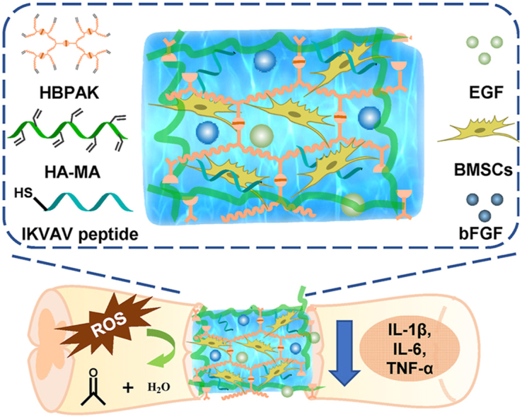
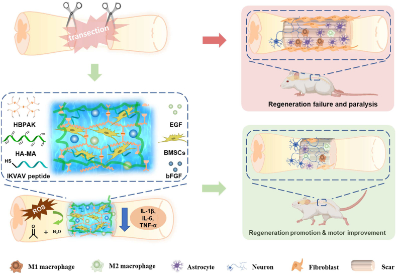
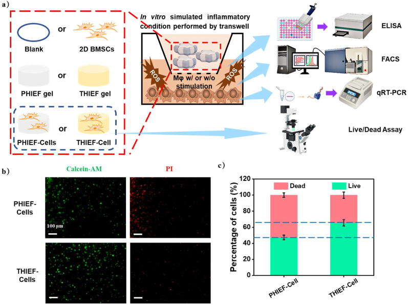
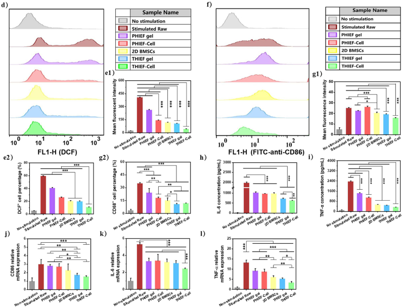
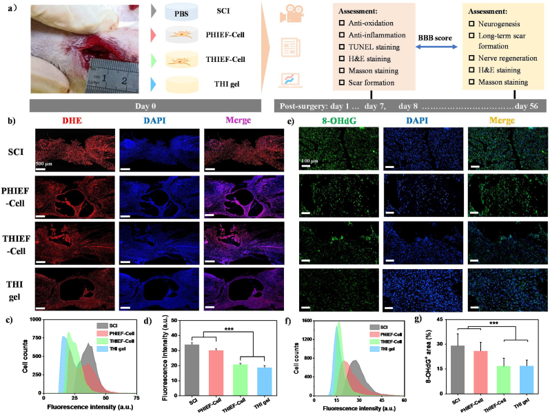
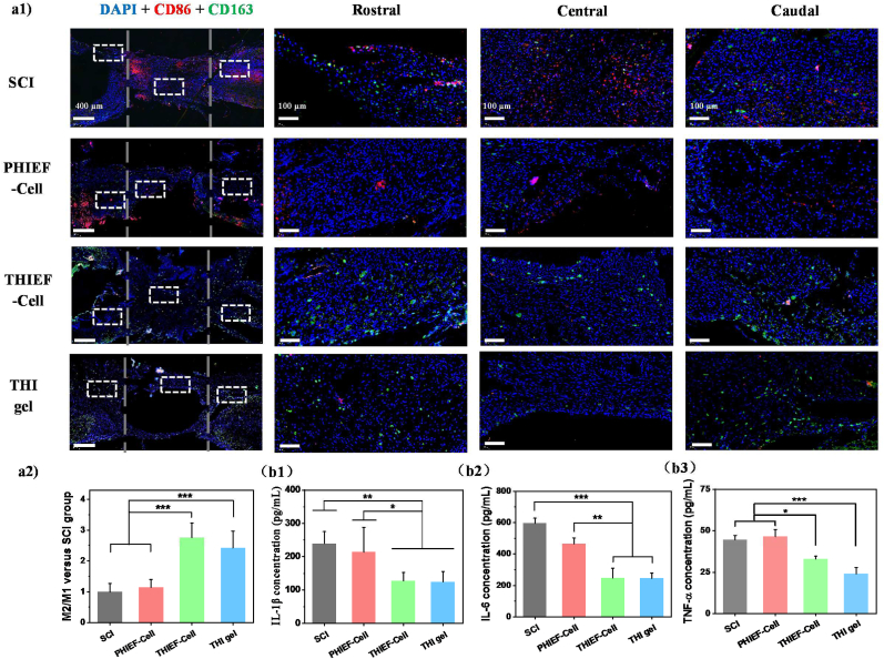
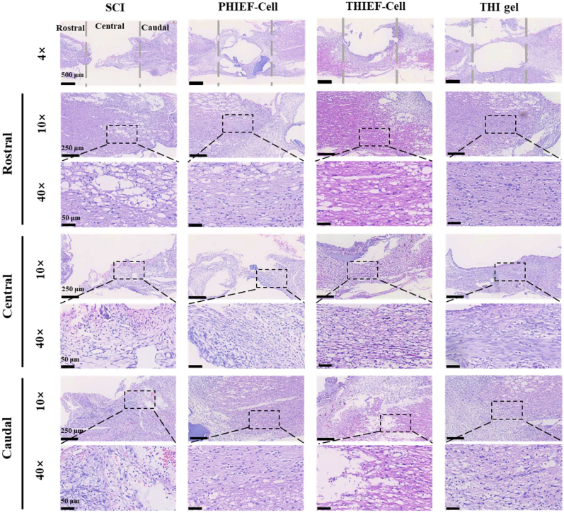
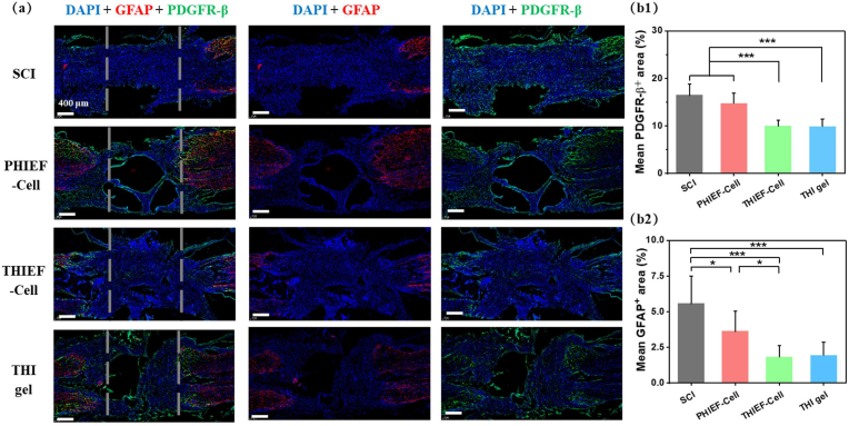
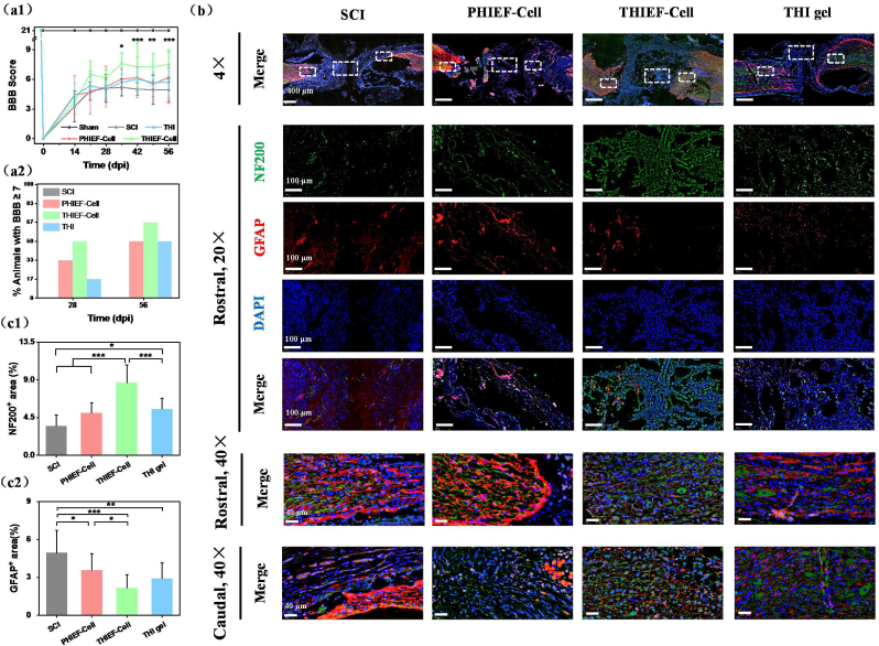
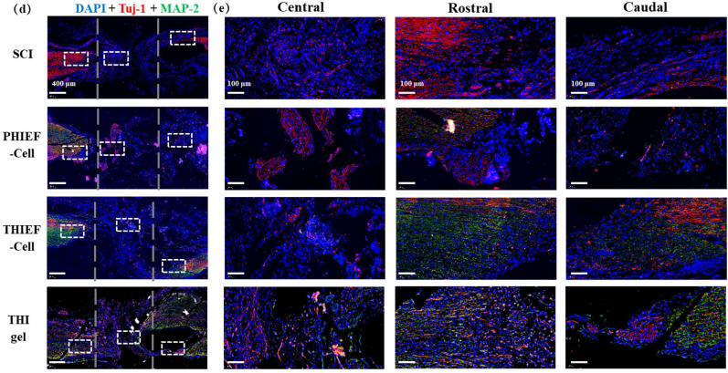
Spinal cord injury (SCI) is an overwhelming and incurable disabling event accompanied by complicated inflammation-related pathological processes, such as excessive reactive oxygen species (ROS) produced by the infiltrated inflammatory immune cells and released to the extracellular microenvironment, leading to the widespread apoptosis of the neuron cells, glial and oligodendroctyes. In this study, a thioketal-containing and ROS-scavenging hydrogel was prepared for encapsulation of the bone marrow derived mesenchymal stem cells (BMSCs), which promoted the neurogenesis and axon regeneration by scavenging the overproduced ROS and re-building a regenerative microenvironment. The hydrogel could effectively encapsulate BMSCs, and played a remarkable neuroprotective role in vivo by reducing the production of endogenous ROS, attenuating ROS-mediated oxidative damage and downregulating the inflammatory cytokines such as interleukin-1 beta (IL-1β), interleukin-6 (IL-6) and tumor necrosis factor-alpha (TNF-α), resulting in a reduced cell apoptosis in the spinal cord tissue. The BMSCs-encapsulated ROS-scavenging hydrogel also reduced the scar formation, and improved the neurogenesis of the spinal cord tissue, and thus distinctly enhanced the motor functional recovery of SCI rats. Our work provides a combinational strategy against ROS-mediated oxidative stress, with potential applications not only in SCI, but also in other central nervous system diseases with similar pathological conditions.

Keywords: Anti-oxidation; Axon regeneration; BMSCs; ROS scavenging; Spinal cord injury (SCI).


Conflict of interest statement

The authors declare that they have no known competing financial interests or personal relationships that could have appeared to influence the work reported in this paper.

Figures

Image 1
Graphical abstract
Scheme 1
Scheme 1
Schematic illustration of the BMSCs-encapsulated ROS-scavenging hydrogel for spinal cord injury treatment. After SCI, the BMSCs-encapsulated ROS-scavenging hydrogel (THIEF-Cell) can eliminate the over-produced ROS to achieve better neuron proration, and significantly improve neurogenesis and motor recovery while attenuate scar formation.
Fig. 1
Fig. 1
Properties of different types of hydrogels. (a) Oscillatory tests as a function of (a1) time and (a2) frequency, and (a3) G′ and G″ measured thereof. The strain and temperature were kept as 1% and 37 °C for the time and frequency sweep tests, respectively, and the frequency was maintained at 6.28 rad/s for the time sweep. (b) Degradation of THI hydrogel and PHI hydrogel in 200 μM H2O2/PBS and pure PBS at 37 °C. (c,d) ROS-scavenging properties of HBPAK-containing THI hydrogels and their elimination efficacy, including (c1)·O2 scavenging properties and (c2) its quantitative analysis of elimination efficiency, (d1) H2O2 elimination properties by using H2O2-KI assessment and (d2) its quantitative analysis of elimination efficiency. The HBPAK, PEGDA, HA-MA and LAP concentrations were kept at 10% w/v, 0.6% w/v, 1% w/v, and 0.05 w/v, respectively to prepare the corresponding hydrogels.
Fig. 2
Fig. 2
Cytotoxicity and cytocompatibility of different hydrogels. Calcein AM and propidium iodide (PI) Live/Dead staining on BMSCs encapsulated in different hydrogels with a density of 8 × 106 per mL post culture for (a) 1 day and 3 days. Quantitative analysis of (b) cell numbers and (c) living cell percentages.
Fig. 3
Fig. 3
In vitro simulated environment of inflammation and oxidative damage performed by co-culturing in a Transwell chamber, including protection of ROS-scavenging hydrogel towards the encapsulated BMSCs. (a) Schematic illustration of the simulated inflammation model and co-culture of cells at different conditions in vitro, including hydrogels with or without BMSCs encapsulation, pure 2D culture of BMSCs in the upper chamber and the stimulated Raw 264.7 in the lower chamber, and their characterization methods. (b) Calcein AM and PI Live/Dead staining on encapsulated BMSCs in THIEF and PHIEF hydrogels for 1 day to evaluate the viability in oxidative condition. (c) quantitative analysis of live cells. (d–g) Flow cytometry and histograms of (d, e) DCF and (f, g) FITC fluorescence intensity of the DCFH-DA or FITC-anti-mouse-CD86 stained Raw 264.7, including (e1) mean DCF fluorescence intensity, (e2) DCF+ cell percentage, (g1) mean FITC fluorescence intensity and (g2) CD86+ cell percentage. (h,i) Expression levels of pro-inflammatory (h) IL-6 and (i) TNF-α tested by ELISA assay in the supernatant of Raw 264.7 medium from each group collected at 1 day after co-culture. (j–l) Transcription levels of pro-inflammatory (j) CD86, (k) IL-6 and (l) TNF-α tested by qRT-PCR assay in the harvested Raw 264.7 at 1 day after co-culture. Data are presented as mean ± SD (n = 4).
Fig. 3
Fig. 3
In vitro simulated environment of inflammation and oxidative damage performed by co-culturing in a Transwell chamber, including protection of ROS-scavenging hydrogel towards the encapsulated BMSCs. (a) Schematic illustration of the simulated inflammation model and co-culture of cells at different conditions in vitro, including hydrogels with or without BMSCs encapsulation, pure 2D culture of BMSCs in the upper chamber and the stimulated Raw 264.7 in the lower chamber, and their characterization methods. (b) Calcein AM and PI Live/Dead staining on encapsulated BMSCs in THIEF and PHIEF hydrogels for 1 day to evaluate the viability in oxidative condition. (c) quantitative analysis of live cells. (d–g) Flow cytometry and histograms of (d, e) DCF and (f, g) FITC fluorescence intensity of the DCFH-DA or FITC-anti-mouse-CD86 stained Raw 264.7, including (e1) mean DCF fluorescence intensity, (e2) DCF+ cell percentage, (g1) mean FITC fluorescence intensity and (g2) CD86+ cell percentage. (h,i) Expression levels of pro-inflammatory (h) IL-6 and (i) TNF-α tested by ELISA assay in the supernatant of Raw 264.7 medium from each group collected at 1 day after co-culture. (j–l) Transcription levels of pro-inflammatory (j) CD86, (k) IL-6 and (l) TNF-α tested by qRT-PCR assay in the harvested Raw 264.7 at 1 day after co-culture. Data are presented as mean ± SD (n = 4).
Fig. 4
Fig. 4
(a) Schematic illustration to show the implantation of BMSC-encapsulated non ROS-responsive hydrogels (PHIEF-Cell), BMSC-encapsulated ROS-responsive hydrogels (THIEF-Cell) and ROS-responsive hydrogel (THI hydrogel) into rats with a 2.0 mm spinal cord transection, and analysis of their antioxidation effects post surgery for 7 days. (b∼d) Dihydroethidium (DHE) staining showing the superoxide anion levels at the lesion site in vivo. (b) Corresponding representative micrographs, together with (c) their histograms showing the fluorescence intensity, and (d) the mean fluorescence intensity calculated from 9 fluorescence images. (e∼g) Immunofluorescent staining of oxidative DNA damage products 8-hydroxy-2′- deoxyguanosine (8-OHdG) at the lesion site of the spinal cord tissues, suggesting the effective antioxidation and protective functions of the HBPAK-containing ROS-responsive hydrogel in vivo. Extent of oxidative damage was analyzed in terms of histogram of (f) the fluorescence intensity and (g) mean 8-OHdG positive area calculated from 9 micrographs (size: 1920 × 1080 pixels) from at least 3 sections for each group. Data represent as mean ± SD. ***p < 0.001.
Fig. 5
Fig. 5
Assessment of anti-inflammatory properties of BMSC-encapsulated ROS-responsive hydrogel post implantation for 7 days in rats with 2.0 mm spinal cord transection. (a1) CD86 (M1 marker) and CD163 (M2 marker) double immunofluorescence staining images, and magnified images in the white box regions, respectively. (a2) M2/M1 ratio determined at the lesion sites. (b) ELISA assay of inflammatory cytokines collected at tissue supernatants for (b1) IL-1β, (b2) IL-6 and (b3) TNF-α. (c) H&E staining images of rat spinal cord tissues at the lesion sites post treatment for 7 days in vivo. The distance between two grey dashed lines in the first panel outline the 2.0 mm gap. *p < 0.05, **p < 0.01, ***p < 0.001.
Fig. 5
Fig. 5
Assessment of anti-inflammatory properties of BMSC-encapsulated ROS-responsive hydrogel post implantation for 7 days in rats with 2.0 mm spinal cord transection. (a1) CD86 (M1 marker) and CD163 (M2 marker) double immunofluorescence staining images, and magnified images in the white box regions, respectively. (a2) M2/M1 ratio determined at the lesion sites. (b) ELISA assay of inflammatory cytokines collected at tissue supernatants for (b1) IL-1β, (b2) IL-6 and (b3) TNF-α. (c) H&E staining images of rat spinal cord tissues at the lesion sites post treatment for 7 days in vivo. The distance between two grey dashed lines in the first panel outline the 2.0 mm gap. *p < 0.05, **p < 0.01, ***p < 0.001.
Fig. 6
Fig. 6
Assessment of formation of both fibrotic scar and glial scar. (a) PDGFR-β (platelet-derived growth factor receptor-β, fibrotic scar marker) and GFAP (glial fibrillary acidic protein, glial scar marker) double immunofluorescence staining of spinal cord tissues at the lesion sites. (b) quantitative analysis of scaring levels by (b1) PDGFR-β+-positive area and (b2) GFAP+-positive area. The dashed lines outline the 2 mm lesion sites.
Fig. 7
Fig. 7
Assessment of motor functional recovery and axon regeneration of spinal cord transected rats after treatment by BMSC-encapsulated hydrogels at 56 days post surgery. (a1) Functional recovery evaluated through Basso-Beattie-Bresnahan (BBB) score. (a2) Analysis of % of animals with a BBB score ≥7 on day 28 and 56 post surgery, suggesting the function restoration in the presence of BMSCs–containing ROS responsive hydrogel. (b) NF200 (neurofilament-200, marker of axon) and GFAP double immunofluorescence staining of nerve fibers and glial scars. The magnified images show the specific areas in the white boxes, and nerve structure in rostral and caudal regions. Quantitative analysis of (c1) NF200 positive and (c2) GFAP positive areas, showing a better neuron preservation and axon regeneration effect of BMSCs-encapsulated ROS-scavenging hydrogel. (d) Tuj-1 (tubulin-III, marker of neurogenesis) and MAP-2 (microtubule-associated protein-2, marker of mature neurons) immunofluorescence double staining images of nerve structure. (e) The magnified images show specific areas in the white boxes. The dashed lines outline the 2 mm transection. *p < 0.05, **p < 0.01, ***p < 0.001.
Fig. 7
Fig. 7
Assessment of motor functional recovery and axon regeneration of spinal cord transected rats after treatment by BMSC-encapsulated hydrogels at 56 days post surgery. (a1) Functional recovery evaluated through Basso-Beattie-Bresnahan (BBB) score. (a2) Analysis of % of animals with a BBB score ≥7 on day 28 and 56 post surgery, suggesting the function restoration in the presence of BMSCs–containing ROS responsive hydrogel. (b) NF200 (neurofilament-200, marker of axon) and GFAP double immunofluorescence staining of nerve fibers and glial scars. The magnified images show the specific areas in the white boxes, and nerve structure in rostral and caudal regions. Quantitative analysis of (c1) NF200 positive and (c2) GFAP positive areas, showing a better neuron preservation and axon regeneration effect of BMSCs-encapsulated ROS-scavenging hydrogel. (d) Tuj-1 (tubulin-III, marker of neurogenesis) and MAP-2 (microtubule-associated protein-2, marker of mature neurons) immunofluorescence double staining images of nerve structure. (e) The magnified images show specific areas in the white boxes. The dashed lines outline the 2 mm transection. *p < 0.05, **p < 0.01, ***p < 0.001.

References

    1. Cowan H., Lakra C., Desai M. Autonomic dysreflexia in spinal cord injury. BMJ Br. Med. J. (Clin. Res. Ed.) 2020;371:m3596. - PubMed
    1. Jiang W., Li M., He F., Zhu L. Inhibition of NLRP3 inflammasome attenuates spinal cord injury-induced lung injury in mice. J. Cell. Physiol. 2019;234(5):6012–6022. - PubMed
    1. James S.L., Theadom A., Ellenbogen R.G., Bannick M.S., Montjoy-Venning W., Lucchesi L.R., Abbasi N., Abdulkader R., Abraha H.N., Adsuar J.C., Afarideh M., Agrawal S., et al. Global, regional, and national burden of traumatic brain injury and spinal cord injury, 1990–2016: a systematic analysis for the Global Burden of Disease Study 2016. Lancet Neurol. 2019;18(1):56–87. - PMC - PubMed
    1. Hutson T.H., Di Giovanni S. The translational landscape in spinal cord injury: focus on neuroplasticity and regeneration. Nat. Rev. Neurol. 2019;15(12):732–745. - PubMed
    1. Badhiwala J.H., Wilson J.R., Witiw C.D., Harrop J.S., Vaccaro A.R., Aarabi B., Grossman R.G., Geisler F.H., Fehlings M.G. The influence of timing of surgical decompression for acute spinal cord injury: a pooled analysis of individual patient data. Lancet Neurol. 2021;20(2):117–126. - PubMed

LinkOut - more resources