. 2022 May 13:19:678-689.
doi: 10.1016/j.bioactmat.2022.05.008. eCollection 2023 Jan.

An immunomodulatory polypeptide hydrogel for osteochondral defect repair

Affiliations

An immunomodulatory polypeptide hydrogel for osteochondral defect repair

Meng Yang et al. Bioact Mater. .

Abstract

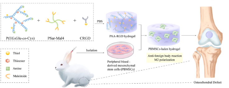
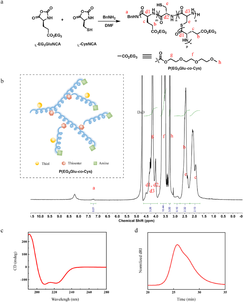
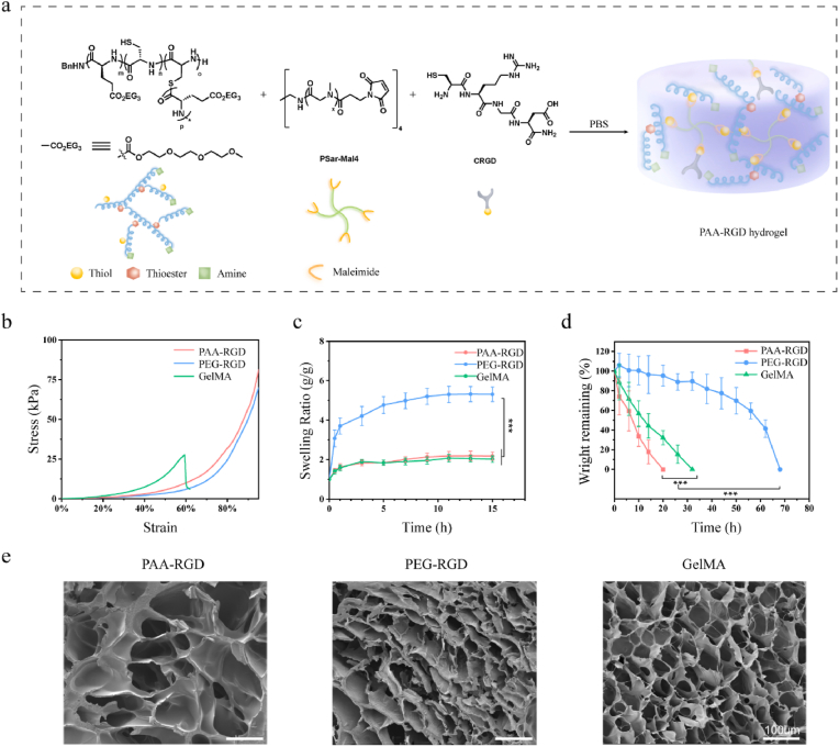
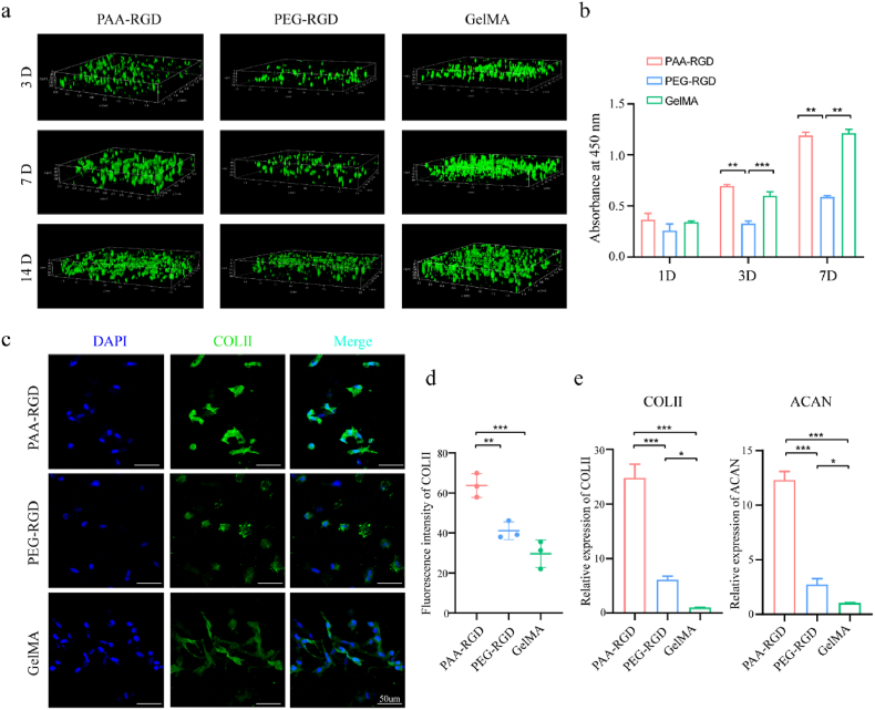
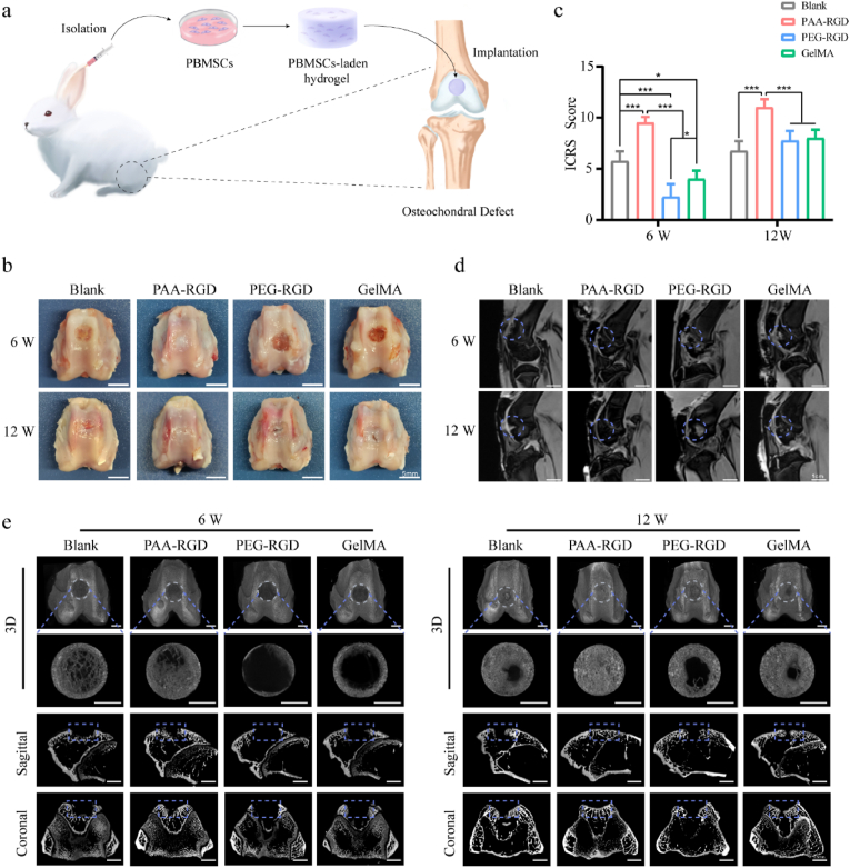
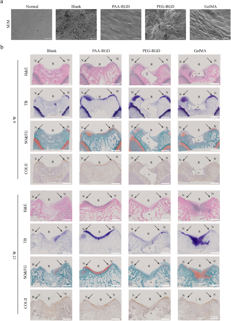
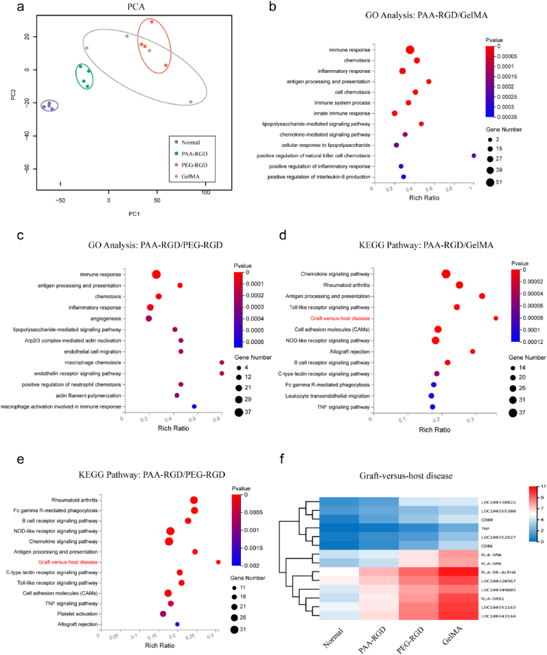
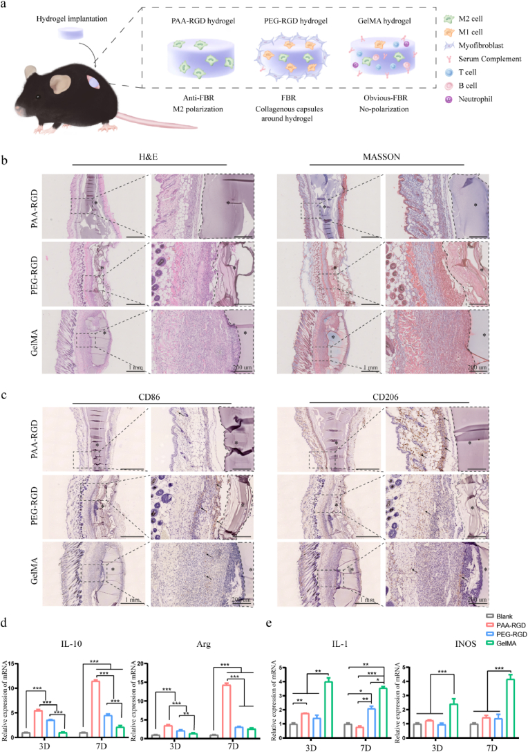
Osteochondral injury is a common and frequent orthopedic disease that can lead to more serious degenerative joint disease. Tissue engineering is a promising modality for osteochondral repair, but the implanted scaffolds are often immunogenic and can induce unwanted foreign body reaction (FBR). Here, we prepare a polypept(o)ide-based PAA-RGD hydrogel using a novel thiol/thioester dual-functionalized hyperbranched polypeptide P(EG3Glu-co-Cys) and maleimide-functionalized polysarcosine under biologically benign conditions. The PAA-RGD hydrogel shows suitable biodegradability, excellent biocompatibility, and low immunogenicity, which together lead to optimal performance for osteochondral repair in New Zealand white rabbits even at the early stage of implantation. Further in vitro and in vivo mechanistic studies corroborate the immunomodulatory role of the PAA-RGD hydrogel, which induces minimum FBR responses and a high level of polarization of macrophages into the immunosuppressive M2 subtypes. These findings demonstrate the promising potential of the PAA-RGD hydrogel for osteochondral regeneration and highlight the importance of immunomodulation. The results may inspire the development of PAA-based materials for not only osteochondral defect repair but also various other tissue engineering and bio-implantation applications.

Keywords: Foreign-body reaction; Immunoregulation; Mesenchymal stem cells; Osteochondral regeneration; Polypeptide hydrogel.


Conflict of interest statement

The authors declare that they have no known competing financial interests or personal relationships that could have appeared to influence the work reported in this paper.

Figures

Image 1
Graphical abstract
Fig. 1
Fig. 1
Synthesis and characterization of P(EG3Glu-co-Cys). (a) Scheme for the synthesis of P(EG3Glu-co-Cys). (b) 1H NMR spectrum (400 M, D2O). (c) CD spectrum in H2O (0.5 mg/mL). (d) SEC trace in DMF.
Fig. 2
Fig. 2
Preparation and characterization of PAA-RGD, PEG-RGD, and GelMA hydrogels. (a) Cartoon illustration of the PAA-RGD hydrogel formation. (b) Strain-stress curves of the hydrogels in the compression study. (c) Swelling curves of the hydrogels (P < 0.001). (d) Protease K-induced degradation rate of PAA-RGD, PEG-RGD, or GelMA hydrogels in vitro (P < 0.001). (e) Representative SEM images of the hydrogels. Scale bars, 100 μm. The polymer content was fixed at 10 wt% for all of the hydrogels. Statistical significance was calculated using one-way ANOVA with Tukey's post-hoc test. ***P < 0.001.
Fig. 3
Fig. 3
The proliferation and chondrogenic differentiation of PB-MSCs in PAA-RGD, PEG-RGD, and GelMA hydrogels. (a) Cell proliferation and Live/Dead staining in 3D plots after 3, 7, and 14 days of culture live cells: green; dead cells: red). (b) Cell proliferation determined by CCK8 assay. (c–d) Immunofluorescence staining (c) and quantification (d) of COL-II expression in PB-MSCs-encapsulating hydrogels on Day 14 following chondrogenic induction; scale bar = 50 μm; (Relative fluorescence intensity was quantified using Image J software). (e) The relative mRNA expression of chondrogenic genes (COL-II, and ACAN) on Day 7 after chondrogenic induction. Data are presented as the mean ± S.D. (n ≥ 3). Statistical significance was calculated using one-way ANOVA with Tukey's post-hoc test. *P < 0.05, **P < 0.01, ***P < 0.001.
Fig. 4
Fig. 4
The repair efficacy of osteochondral defects by PBMSCs-encapsulated hydrogels in vivo. (a) The operation flowchart of PBMSCs isolation, encapsulation, and hydrogel implantation into osteochondral defects. (b) The macroscopic appearance of the repaired joint; scale bar = 5 mm. (c) ICRS macroscopic assessment scores in osteochondral repair. Data are presented as the mean ± S.D. (n = 4). Statistical significance was calculated using two-way ANOVA with Tukey's post-hoc test. *P < 0.05, ***P < 0.001. (d) The MRI scan of the repaired knee joints; scale bars = 1 cm. (e) Representative micro-CT images of subchondral bone repair at 6 and 12 weeks after treatment; scale bar = 2.5 mm.
Fig. 5
Fig. 5
Morphology, histology, and immunohistochemistry of the repaired cartilages. (a) SEM images of representative repaired cartilage surfaces 12 weeks after treatments. scale bar = 40 μm. (b) H&E, Toluidine Blue (purple), Safranin O-Fast Green (orange & green), and immunohistochemistry staining showing the COL-II regeneration (brown); scale bar = 2 mm, * = the nondegraded part of the hydorgel, N = normal cartilage, R = repaired tissues, the arrows indicated the interfaces of the normal cartilages and repaired tissues.
Fig. 6
Fig. 6
RNA sequencing of the repaired articular cartilages at week 12. (a) Principal component analysis (PCA) of the cartilage transcriptome from the normal, PAA-RGD, PEG-RGD, and GelMA groups. (b–c) Gene ontology (GO) pathway analysis of genes downregulated in the PAA-RGD group as relative to the GelMA (b) and PEG-RGD (c) groups. (d–e) KEGG pathway analysis of genes downregulated in the PAA-RGD group as relative to the GelMA (d) and PEG-RGD (e) groups. (f) Hierarchical clustering analysis using differentially expressed genes implied downregulated graft-versus-host disease pathways for the PAA-RGD group.
Fig. 7
Fig. 7
FBR and macrophage polarization of various hydrogels implanted in mice and RAW264.7 cells. (a) Cartoon illustration of FBR and macrophages polarization in C57/BL6 mice induced by the subcutaneously implanted hydrogels. (b) HE and MASSON staining were used to evaluate the inflammatory response and collagen encapsulation density after 14 days after implantation. Scale bars, 1 mm, 200 μm; * = hydrogels. (c) Immunohistochemical staining to assess the phenotype of macrophages infiltrated into hydrogels-tissues interfaces on Day14 after implantation. Positive staining is observed as a brown color (Representative positive cells indicated by arrows), while all nucleus stained with hematoxylin show blue color. Scale bars, 1 mm, 200 μm; * = hydrogels. (d–e) The mRNA levels of the representative M2 macrophages biomarkers IL-10 and Arg (d) and the characteristic M1 macrophages biomarkers IL-1 and iNOS (e). RAW 264.7 cells were treated with various hydrogels for 3 and 7 days before harvesting and analyzed by qRT-PCR. Data are presented as the mean ± S.D. (n ≥ 3). Statistical significance was calculated using two-way ANOVA with Tukey's post-hoc test. *P < 0.05, **P < 0.01, ***P < 0.001.

References

    1. Wei W., Dai H. Articular cartilage and osteochondral tissue engineering techniques: recent advances and challenges. Bioact. Mater. 2021;6:4830–4855. doi: 10.1016/j.bioactmat.2021.05.011. - DOI - PMC - PubMed
    1. Zha K., Li X., Yang Z., Tian G., Sun Z., Sui X., Dai Y., Liu S., Guo Q. Heterogeneity of mesenchymal stem cells in cartilage regeneration: from characterization to application. npj Regener. Med. 2021;6:14. doi: 10.1038/s41536-021-00122-6. - DOI - PMC - PubMed
    1. Xiang Y., Bunpetch V., Zhou W., Ouyang H. Optimization strategies for ACI: a step-chronicle review. J. Orthop Translat. 2019;17:3–14. doi: 10.1016/j.jot.2018.12.005. - DOI - PMC - PubMed
    1. Kunze K.N., Manzi J.S., Wright-Chisem J., Ramkumar P., Nwachukwu B.U., Williams R.R. Risk factors for failure after osteochondral allograft transplantation of the knee: a systematic review and exploratory meta-analysis. Am. J. Sports Med. 2022 doi: 10.1177/03635465211063901. 3635465211063901. - DOI - PubMed
    1. Cao H., Wang X., Chen M., Liu Y., Cui X., Liang J., Wang Q., Fan Y., Zhang X. Childhood cartilage ECM enhances the chondrogenesis of endogenous cells and subchondral bone repair of the unidirectional collagen-dECM scaffolds in combination with microfracture. ACS Appl. Mater. Interfaces. 2021;13:57043–57057. doi: 10.1021/acsami.1c19447. - DOI - PubMed

LinkOut - more resources